4
views
0
recommends
+1 Recommend
0 collections
    0
    shares
      • Record: found
      • Abstract: found
      • Article: found
      Is Open Access

      Reversing the miRNA -5p/-3p stoichiometry reveals physiological roles and targets of miR-140 miRNAs

      research-article

      Read this article at

      Bookmark
          There is no author summary for this article yet. Authors can add summaries to their articles on ScienceOpen to make them more accessible to a non-specialist audience.

          Abstract

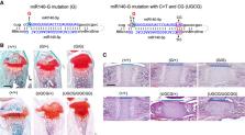
          The chondrocyte-specific miR-140 miRNAs are necessary for normal endochondral bone growth in mice. miR-140 deficiency causes dwarfism and craniofacial deformity. However, the physiologically important targets of miR-140 miRNAs are still unclear. The miR-140 gene ( Mir140) encodes three chondrocyte-specific microRNAs, miR-140-5p, derived from the 5′ strand of primary miR-140, and miR140-3p.1 and -3p.2, derived from the 3′ strand of primary miR-140. miR-140-3p miRNAs are 10 times more abundant than miR-140-5p likely due to the nonpreferential loading of miR-140-5p to Argonaute proteins. To differentiate the role of miR-140-5p and -3p miRNAs in endochondral bone development, two distinct mouse models, miR140-C > T, in which the first nucleotide of miR-140-5p was altered from cytosine to uridine, and miR140-CG, where the first two nucleotides of miR-140-3p were changed to cytosine and guanine, were created. These changes are expected to alter Argonaute protein loading preference of -5p and -3p to increase -5p loading and decrease -3p loading without changing the function of miR140-5p. These models presented a mild delay in epiphyseal development with delayed chondrocyte maturation. Using RNA-sequencing analysis of the two models, direct targets of miR140-5p, including Wnt11, were identified. Disruption of the predicted miR140-5p binding site in the 3′ untranslated region of Wnt11 was shown to increase Wnt11 mRNA expression and caused a modest acceleration of epiphyseal development. These results show that the relative abundance of miRNA-5p and -3p can be altered by changing the first nucleotide of miRNAs in vivo, and this method can be useful to identify physiologically important miRNA targets.

          Related collections

          Most cited references29

          • Record: found
          • Abstract: found
          • Article: found
          Is Open Access

          Predicting effective microRNA target sites in mammalian mRNAs

          MicroRNA targets are often recognized through pairing between the miRNA seed region and complementary sites within target mRNAs, but not all of these canonical sites are equally effective, and both computational and in vivo UV-crosslinking approaches suggest that many mRNAs are targeted through non-canonical interactions. Here, we show that recently reported non-canonical sites do not mediate repression despite binding the miRNA, which indicates that the vast majority of functional sites are canonical. Accordingly, we developed an improved quantitative model of canonical targeting, using a compendium of experimental datasets that we pre-processed to minimize confounding biases. This model, which considers site type and another 14 features to predict the most effectively targeted mRNAs, performed significantly better than existing models and was as informative as the best high-throughput in vivo crosslinking approaches. It drives the latest version of TargetScan (v7.0; targetscan.org), thereby providing a valuable resource for placing miRNAs into gene-regulatory networks. DOI: http://dx.doi.org/10.7554/eLife.05005.001
            Bookmark
            • Record: found
            • Abstract: found
            • Article: not found

            Metazoan MicroRNAs

            MicroRNAs (miRNAs) are ∼22 nt RNAs that direct posttranscriptional repression of mRNA targets in diverse eukaryotic lineages. In humans and other mammals, these small RNAs help sculpt the expression of most mRNAs. This article reviews advances in our understanding of the defining features of metazoan miRNAs and their biogenesis, genomics, and evolution. It then reviews how metazoan miRNAs are regulated, how they recognize and cause repression of their targets, and the biological functions of this repression, with a compilation of knockout phenotypes that shows that important biological functions have been identified for most of the broadly conserved miRNAs of mammals.
              Bookmark
              • Record: found
              • Abstract: found
              • Article: not found

              Regulation of microRNA function in animals

              Since their serendipitous discovery in nematodes, microRNAs (miRNAs) have emerged as key regulators of biological processes in animals. These small RNAs form complex regulatory networks in cell development, differentiation and homeostasis. Deregulation of miRNA function is associated with an increasing number of human diseases, particularly cancer. Recent discoveries have expanded our understanding of how miRNAs are regulated. Here we review the mechanisms that modulate miRNA activity, their stability and their localization through alternative processing, sequence editing, post-translational modifications of Argonaute proteins, viral factors, transport from the cytoplasm and regulation of miRNA–target interactions. We conclude by discussing intriguing open questions to be answered by future research.
                Bookmark

                Author and article information

                Journal
                RNA
                RNA
                RNA
                RNA
                Cold Spring Harbor Laboratory Press
                1355-8382
                1469-9001
                June 2022
                : 28
                : 6
                : 854-864
                Affiliations
                [1 ]Endocrine Unit, Massachusetts General Hospital, Boston, Massachusetts 02114, USA
                [2 ]Harvard Medical School, Boston, Massachusetts 02114, USA
                Author notes
                Article
                9509184 RA
                10.1261/rna.079013.121
                9074898
                35332065
                e17a1eea-2013-461a-8ca1-ee31a1a3db7a
                © 2022 Young et al.; Published by Cold Spring Harbor Laboratory Press for the RNA Society

                This article is distributed exclusively by the RNA Society for the first 12 months after the full-issue publication date (see http://rnajournal.cshlp.org/site/misc/terms.xhtml). After 12 months, it is available under a Creative Commons License (Attribution-NonCommercial 4.0 International), as described at http://creativecommons.org/licenses/by-nc/4.0/.

                History
                : 8 October 2021
                : 10 March 2022
                Page count
                Pages: 11
                Funding
                Funded by: Center for Skeletal Research
                Award ID: P30AR075042
                Funded by: National Institutes of Health (NIH) , doi 10.13039/100000002;
                Award ID: AR056645
                Categories
                Article

                microrna,mir-140-5p,mir-140-3p,mice,chondrocyte
                microrna, mir-140-5p, mir-140-3p, mice, chondrocyte

                Comments

                Comment on this article