- Record: found
- Abstract: found
- Article: not found
Exome sequencing identifies ACSF3 as the cause of Combined Malonic and Methylmalonic Aciduria
research-article
Jennifer L. Sloan
1 ,
Jennifer J. Johnston
2 ,
Irini Manoli
1 ,
Randy J. Chandler
1
,
3 ,
Caitlin Krause
2 ,
Nuria Carrillo-Carrasco
1 ,
Suma D. Chandrasekaran
1 ,
Justin R. Sysol
1 ,
Kevin O’Brien
4 ,
Natalie S. Hauser
1 ,
Julie C. Sapp
2 ,
Heidi M. Dorward
4 ,
Marjan Huizing
4 ,
NIH Intramural Sequencing Center Group
5 ,
Bruce A. Barshop
6 ,
Susan A. Berry
7 ,
Philip M. James
8 ,
Neena L. Champaigne
9 ,
Pascale de Lonlay
10 ,
Vassilli Valayannopoulos
10 ,
Michael D. Geschwind
11 ,
Dimitar K. Gavrilov
12 ,
William L. Nyhan
6 ,
Leslie G. Biesecker
2 ,
Charles P. Venditti
1
14 August 2011
Read this article at

There is no author summary for this article yet. Authors can add summaries to their articles on ScienceOpen to make them more accessible to a non-specialist audience.
Abstract
We used whole exome sequencing of a single patient with combined malonic and methylmalonic
aciduria (CMAMMA) to identify mutations in ACSF3, a putative malonyl-CoA and methylmalonyl-CoA
synthetase (MCS). Follow-up sequencing of eight additional patients, including an
individual who was diagnosed after mining an exome database as well as an affected
canine, showed pathogenic mutations. ACSF3 mutant alleles occur with a minor allele
frequency (MAF) of 0.0058 in ~1,000 control individuals predicting a CMAMMA population
incidence ~ 1:30,000. CMAMMA is the first human disorder caused by mutations in a
member of the acyl-CoA synthetase family, a diverse group of evolutionarily conserved
proteins, and may emerge as one of the more common human metabolic disorders.
Methylmalonic acidemias (MMAemias) are heterogeneous disorders that exhibit elevated
methylmalonic acid (MMA) in body fluids. Deficiency of methylmalonyl-CoA mutase (MUT)
or the enzymes (MMAA, MMAB, MMADHC) that synthesize 5′-adenosylcobalamin comprise
most disease subtypes. Some patients have atypical forms of MMAemia, e.g., combined
malonic and methylmalonic aciduria (CMAMMA) that lack enzymatic and molecular definition.
CMAMMA was first reported in a child with immunodeficiency, failure to thrive, seizures,
increased urinary MMA compared to malonic acid (MA) and normal malonyl-CoA decarboxylase
activity
1
. A Labrador retriever with similar biochemical features and neurodegeneration has
also been described
2
.
To determine the cause of CMAMMA, we took a multifaceted approach that included exome
and candidate gene sequencing in nine patients, identification of the canine orthologue
and mutation analysis in an affected dog, and a novel strategy of hypothesis-generating
clinical research in an exome cohort
3
. We establish ACSF3 mutations as the cause of CMAMMA and describe the first disease
association with a member of the acyl-CoA synthetase (ACS) family, enzymes that activate
fatty acids for intermediary metabolism
4
.
Nine subjects with CMAMMA participated and six were evaluated at the NIH. The age
of diagnosis and symptoms were variable (Table 1). After uneventful early decades,
four patients were diagnosed in adulthood with neurological manifestations (seizures,
memory problems, psychiatric disease, and/or cognitive decline) without vitamin B12
deficiency. Five subjects presented during childhood with symptoms suggestive of an
intermediary metabolic disorder (coma, ketoacidosis, hypoglycemia, failure to thrive,
elevated transaminases, microcephaly, dystonia, axial hypotonia, and/or developmental
delay).
Methylmalonic and malonic aciduria with urinary MMA/MA >5 was present in seven of
nine affecteds (Table 1). Serum MMA was elevated but serum B12 levels, acylcarnitines,
and total homocysteine were normal, as were malonyl-CoA decarboxylase activity, 1-C14-propionate
incorporation, malonyl-CoA decarboxylase (MLYCD) genetic testing, and sequencing of
known MMAemia genes (Table 1). Plasma MA was measured by GC/MS in six patients and
was also markedly elevated (Table 1). We conclude that these subjects all have CMAMMA,
which is distinct from other forms of MMAemia.
For Subject 1, we sequenced target-selected libraries in the paired-end 101 bp configuration,
yielding 114,467 variant genotypes. We used genetic filters for homozygosity or compound
heterozygosity. We included nonsynonymous, splice, frameshifting, and nonsense variants
as potential mutations but excluded dbSNP variants. We used control exome data
3
to exclude homozygous variants or variants with >10% frequency (Supplementary Table
1 and Online Methods).
The filtering strategy yielded 12 genes, from which we selected for further evaluation
ACSF3, an orphan member of the acyl-CoA synthetase family, based on its putative function
and predicted mitochondrial localization. We found three ACSF3 exome variants in Subject
1 (c.1385A>C p.Lys462Thr, c.del1394_1411, p.Gln465_Gly470del, and c.1627C>T, p.Arg558Trp)
and confirmed them by Sanger sequencing (Table 1, Supplementary Figure 1, Supplementary
Table 2). The variants p.Lys462Thr and p.Gln465_Gly470del were in trans with p.Arg558Trp
based on parental genotypes and segregated in two unaffected siblings who, like their
parents, had normal serum MMA levels. We sequenced ACSF3 exons in seven additional
patients with CMAMMA; six had ACSF3 variations (Table 1, Supplementary Figure 1).
One patient had no damaging mutations detected.
Next, we identified a putative canine ACSF3 orthologue and sequenced DNA from the
CMAMMA Labrador retriever, this showed a homozygous alteration (c.1288G>A, p.Gly430Ser;
orthologous to human p.Gly480) in a conserved residue (Figure 1, Table 1, Supplementary
Figure 1). This variant was absent in 40 control Labrador DNAs selected for maximum
diversity based on American Kennel Club numbers. Finally, we took a novel approach
to patient discovery by analyzing exome data of 401 individuals ascertained for cardiovascular
phenotypes
3
. We identified a 66 year-old female, apparently homozygous for a c.1411C>T, p.Arg471Trp
ACSF3 variant. She had no known metabolic disease symptoms but reported incontinence
and mild memory problems. Her laboratory evaluation showed 48 μM MMA and 11.3 μM MA
in plasma and 206 mmol/mol Cr MMA and 26.3 mmol/mol Cr MA in urine, and normal serum
B12 levels and acylcarnitines. We did not find mutations in other known MMAemia genes
in her exome (Table 1).
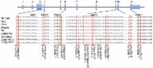
We identified nine missense, one in-frame deletion and one nonsense mutation (Figure
1). Four subjects were apparently homozygous for ACSF3 variants. Although it is possible
that unidentified deletions might play a role in this disorder, this is unlikely for
these individuals (Table 1). Most of the variants resided in the C-terminal half of
ACSF3. Eight out of nine missense mutations and the in-frame deletion were located
in conserved ACS motifs predicted to be involved in AMP binding (Motif I), conformational
change and catalytic function (Motif II), substrate binding (Motifs III, IV), or catalysis
(Motif V)
5
(Figure 1). Western analyses using fibroblasts from Subjects 1–4 and 7 showed the
presence of cross-reactive ACSF3 (Supplementary Figure 2). Fibroblasts from Subjects
1–4 produced 2.4- to 6-fold more MMA than control cells (Figure 2A) after chemical
stimulation. Viral expression of ACSF3, but not GFP (Figure 2B) restored metabolism,
and provided validation of ACSF3 function in a cell culture biochemical assay.
These data establish a candidate gene for CMAMMA using exome sequencing in a single
affected with validation using four approaches. First, seven additional probands harbored
two mutations in ACSF3. Second, an affected dog had a single, unique sequence variant
in the canine ACSF3 orthologue in a conserved residue that was absent in 40 diverse
controls. Third, one patient with two ACSF3 mutations was identified in a cohort of
subjects not ascertained for metabolic disease and had biochemical features of CMAMMA.
Finally, viral complementation of ACSF3 in patient fibroblasts corrected the cellular
metabolic defect. Based on these observations, we conclude that mutations in ACSF3
cause CMAMMA.
The ACSF3 gene is an orphan member of the acyl-coenzyme A synthetase gene family,
enzymes that thioesterify substrates into CoA derivatives, and weakly activated C24:0
fatty acid
4
. The biochemical abnormalities in the patients led us to reassess the possible function
of ACSF3. When we compared human ACSF3 to Bradyrhizobium japonicum malonyl-CoA synthetase
(MCS), a well-characterized enzyme, the proteins were more identical (32%) and similar
(50%) to each other than ACSF3 was to the next closest human ACS family member (ACSM3v1,
28% identity). Phylogenetic analyses rooted human ACSF3 with the MCS enzymes versus
other ACSs (Supplementary Figure 3). To provide preliminary experimental evidence
for predicted function, we examined purified, GST-tagged ACSF3 under MCS assay conditions
and found that the enzyme activated malonate and methylmalonate, but not acetate,
into the respective coenzyme thioesters (Supplementary Table 3). The specific activity
of GST-tagged ACSF3 was higher with malonate as a substrate compared to methylmalonate,
similar to its prokaryotic homologues. Because the first 58 amino acids of ACSF3 are
predicted to encode a mitochondrial leader sequence (Supplementary Figure 4), we performed
immunostaining with fibroblasts overexpressing ACSF3 and a C-terminal GFP-ACSF3 fusion
protein. ACSF3 staining showed a distinct mitochondrial distribution and co-localized
with a mitochondrial antibody (Figure 3). The comparative sequence analysis, enzymatic
data, and subcellular localization lead us to propose that ACSF3 is a mitochondrial
malonyl-CoA and methylmalonyl-CoA synthetase (MCS), an enzyme postulated to catalyze
the first step of intramitochondrial fatty acid synthesis
5
.
The assignment of ACSF3 as an MCS provides a framework to understand the consequences
of the ACSF3 mutations and the metabolic perturbations of CMAMMA. MCS from R. trifolii
and B. japonicum activate malonate and methylmalonate as substrates in vitro
6,7
as does ACSF3 (Supplementary Table 3), suggesting that malfunction of this enzyme
causes accretion of the proximal substrates that manifests as methylmalonic and malonic
aciduria. Site-directed mutagenesis experiments with B. japonicum MCS showed that
p.Glu308Gln abolishes malonate binding
7
. The corresponding human ACSF3 position is the residue mutated in Subject 3, p.Glu359Lys
in Motif III, and predicts that this mutation is likely to effect the Km for malonate.
Arg471 in motif II is nearly invariant in the ACS family
4
and essential for acyl-CoA synthetase activity
8–10
. Therefore, an Arg471 alteration in ACSF3, as in Subjects 5 and 6, likely affects
enzymatic function. Other missense alterations (p.Pro243Leu, p.Thr358Ile, p.Gly430Ser
(dog), p.Arg558Trp) map to conserved residues in B. japonicum and R. leguminosarum
MCS (Figure 1) or to conserved residues in other ACSF3 family members (p.Met198Arg,
p.Lys462Thr), and are likely to be deleterious.
In the ClinSeq™ cohort, there were an additional four participants heterozygous for
ACSF3 variants (p.Glu359Lys n=1, p.Arg558Trp n=3) also found in patients with CMAMMA.
The 1000 genomes dataset (estimated coverage of 629 genomes) there were six individuals
with ACSF3 mutations (p.Glu359Lys n=1, p.Arg558Trp n=5). Combining these data yields
an overall MAF of 0.0058 (95% CI, .0033–.0106) for an estimated disease incidence
of ~1/30,000 (95% CI, 1/9,000 – 1/92,000). We predict that CMAMMA is one of the most
common forms of MMAemia
11
, and perhaps, one of the most common inborn errors of metabolism. Clearly, the spectrum
of symptoms and natural history of this disorder are highly variable and require further
delineation. The identification of an affected using exome sequencing highlights an
interesting and alterative diagnostic approach because CMAMMA is not identified through
routine newborn screening (via elevated propionylcarnitine [C3]). We speculate that
CMAMMA and other metabolic disorders that have escaped early diagnosis could be identified
using genomic techniques.
Online Methods
Subjects
Patient studies were approved by the National Human Genome Research Institute Institutional
Review Board as part of NIH studies: 04-HG-0127 “Clinical and Basic Investigations
of Methylmalonic Acidemia and Related Disorders”(ClinicalTrials identifier: NCT00078078),
10-HG-0065 “Whole Genome Medical Sequencing for Genome Discovery” (ClinicalTrials
identifier: NCT01087320), 07-HG-0002 “ClinSeq™: A Large-Scale Medical Sequencing Clinical
Research Pilot Study” (ClinicalTrials identifier: NCT00410241), and/or 94-HG-0193
“Genetic and Clinical Studies of Congenital Anomaly Syndromes” (ClinicalTrials identifier:
NCT00001404) and were performed in compliance with US 45.CFR.46. Adult participants
and parents of the younger subjects signed written informed consent for their participation.
Patients with CMAMMA were evaluated at the NIH Clinical Center and/or outside records
from referring centers were reviewed. Plasma MMA was determined by liquid chromatography-tandem
mass spectrometry (LC-MS/MS) stable isotope dilution analysis and urine organic acids
were measured by gas chromatography-mass spectrometry (GC/MS) (Mayo Medical Laboratories).
A GC/MS assay was developed to measure MA in patient samples (Gavrilov et al, unpublished).
In brief, D3 methylmalonic and 13C2-malonic acid were added to plasma, serum or urine,
adjusted with NaCl, and acidified. An ethyl acetate extraction was performed and the
organic layer was concentrated under N2 flow. The resulting residue was silylated
with BSTFA + 1% TMCS. The samples were then analyzed by GC/MS in the selected ion
monitoring mode. Plasma samples from 19 anonymous controls with normal MMA levels
were used to develop the reference ranges for MA (mean 0.67 μM ± 0.14, range 0.38
to 0.89 μM).
DNA Isolation
DNA was isolated from all human subjects and the canine controls from whole blood
using the salting out method (Qiagen) following the manufacturer’s instructions. For
the CMAMMA canine sample, DNA was isolated by similar methods from a fibroblast cell
line.
Next-Generation sequencing and variant analysis
Solution hybridization exome capture was carried out using the SureSelect Human All
Exon System (Agilent Technologies). Manufacturer’s protocol version 1.0 compatible
with Illumina paired end sequencing was used, with the exception that DNA fragment
size and quality was measured using a 2% agarose gel. Flow cell preparation and 101
bp paired-end read sequencing were carried out as per protocol for the GAIIx sequencer
(2) (Illumina Inc). A single 101 base pair paired-end lane on a GAIIx flow cell was
used per exome sample to generate sufficient reads to generate the aligned sequence.
Image analyses and base calling on all lanes of data were performed using Illumina
Genome Analyzer Pipeline software (GAPipeline versions 1.4.0 or greater) with default
parameters.
Read mapping, variant calling and annotation
Reads were aligned to a human reference sequence (UCSC assembly hg18, NCBI build 36)
using the package called “efficient large-scale alignment of nucleotide databases”
(ELAND). Reads that align uniquely were grouped into genomic sequence intervals of
about 100 kb, and reads that fail to align were binned with their paired-end mates.
Reads in each bin were subjected to a Smith-Waterman-based local alignment algorithm,
cross_match using the parameters – min score 21 and – mask level 0 to their respective
100 kb genomic sequence. Genotypes were called at all positions where there were high-quality
sequence bases (Phred-like Q20 or greater) using a Bayesian algorithm (Most Probable
Genotype – MPG)
12
.
Filtering Strategy
The filters used in this study included a number of criteria that were implemented
using the VarSifter software program for exome and whole genome data management (Teer
et al., unpublished). The filters for homozygosity or compound heterozygosity in the
proband were used because most metabolic diseases are autosomal recessive and those
for mutation type (nonsynonymous, splice, frameshift, and nonsense) were selected
because they encompass the majority of disease-causing variants. However, these filters
would not detect large deletions, regulatory mutations, or non-canonical splice mutations,
which can account for several percent of causative mutations. We also used an exclusion
of alleles present in dbSNP, reasoning that the causative variants were uncommon and
unlikely to be cataloged there. We used a MAF filter of <10% from a cohort of 258
subjects who were sequenced in our center with similar methodology. We recognized
that this frequency was quite liberal, but used it as a starting point, anticipating
that it would be adjusted should the initial screen not have yielded a plausible candidate.
We reasoned, incorrectly in retrospect, that it was appropriate to filter out variants
that were homozygous in controls, as we assumed that no member of the control cohort
could have this disease.
Sanger Sequence Analysis
Sequence analysis of ACSF3 was performed using standard methods. Sequencing was performed
with v3.1 BigDye terminator cycle sequencing kit (Applied Biosystems) and the ABI
3130 (Applied Biosystems) per the manufacturer’s protocol. Sequence data were compared
with the published ACSF3 sequence (GenBank reference number NM_174917.2) using Sequencher
4.10.1 (Gene Codes Corp.). Nucleotide numbering reflects cDNA numbering with +1 corresponding
to the A of the ATG translation initiation codon in the reference sequence. The initiation
codon is codon 1.
Canine ACSF3
As no canine orthologue for ACSF3 was known, the Dog Genome (UCSC browser, May 2005
build) was used to predict the sequence for canine ACSF3 and primers were designed
to amplify the exonic regions of the gene. Liver cDNA was obtained (Zyagen) and primers
for the predicted dog cDNA were used to amplify the transcript. The dog ACSF3 partial
cDNA sequence has been submitted to GenBank, Accession number JF907588.1. DNA from
forty unrelated Labrador retrievers was obtained (E. Ostrander, NHGRI) and tested
for c.1288G>A, p.Gly430Ser.
Sequence alignment and Bioinformatics
ACSF3 orthologues were identified by BLAST search and through Homologene. Sequence
alignment of the ACSF3 orthologues: human NP_001120686.1, mouse NP_659181.2, dog JF907588.1,
cow NP_001030240.1, rat XP_574249.3, zebrafish XP_690782.2, Xenopus NP_001086314.1,
B. japonicum NP_767149.1 and R. leguminosarum AAC83455.1, was performed by the Clustal
W method in MacVector version 9.0.2. The phylogenetic tree was created in MegAlign
(Lasergene) by the Clustal W method. Similarity was determined by BLAST-P using the
BLOSUM 62 matrix. The mitochondrial leader sequence was predicted using MitoProtII.
Expression of human ACSF3 cDNA in human fibroblast cells
Wild-type ACFS3 cDNA was generated by RT-PCR from total RNA extracted from normal
human liver tissue and sequence validated. This gene was cloned into a Gateway (Invitrogen)
retroviral expression vector, pLenti6/V5-DEST, as recommended by the manufacturer.
The viral constructs express ACSF3 or GFP under the control of the CMV promoter; the
backbone also has a blasticidin cassette driven by the E7 promoter. Human fibroblast
cell lines were transduced with virus containing either the ACSF3 or GFP. The transduced
cells were selected and expanded in DMEM with 5% fetal bovine serum containing 10
μg/ml blasticidin, for selection, prior to propionate loading. ACSF3 with a C-terminal
GFP fusion was cloned into pCMV6 and sequence verified. Control fibroblasts were electroporated
with 3 μg of plasmid DNA using an Amaxa nucleofector electroporator (Amaxa GmbH, Walkersville,
MD). Transfected fibroblasts were grown for 48 hours before immunofluorescence experiments.
MMA production by cultured CMAMMA fibroblasts
A modified chemical stimulation study was performed as described
13
. Six well tissue culture plates were seeded at a density of 2 or 5×10⁵ per well in
high glucose (4 g/L) DMEM supplemented with 10% fetal bovine serum, penicillin streptomycin,
L-glutamine and sodium pyruvate. The next day, the DMEM growth media was removed and
replaced with 1 ml of DMEM growth media containing sodium propionate at a concentration
of 5 mM. After 72 hours the media was collected for GC/MS analysis of MMA.
Western blot analyses
Thirty to forty micrograms of clarified fibroblast extract were analyzed by Western
blot using a rabbit polyclonal anti-ACSF3 (ab100860; Abcam) or mouse monoclonal anti-PDH-E2
(MSP05; MitoSciences) at a dilution of 1:1,500. Mouse monoclonal anti-β-actin (ab8226,
Abcam) was used as a loading control for immunoblotting at a dilution of 1:1,000.
Horseradish peroxidase–conjugated anti-rabbit IgG or anti-mouse IgG (NA934 or NA931;
GE Healthcare Life Sciences) was used as the secondary antibody and was visualized
with chemiluminescence detection (Pierce Biotechnology).
Enzyme Assay
Full-length ACSF3 containing an N-terminal GST fusion, expressed in wheat germ extract,
was obtained from Novus Biologicals and used to assay malonyl- and methylmalonyl-CoA
synthetase activity with a previously described spectrophotometric method
7
. The reaction mixture contained the following components in a volume of 500 μL: 100
mM potassium phosphate buffer (pH 7.0), 8 mM malonate, methylmalonate, or acetate,
2 mM MgCl2, 0.4 mM ATP, 0.2 mM CoA, and 1.43 μg of GST-tagged, purified ACSF3. An
increase in absorbance at 232 nm was used to measure the formation of the thioester
bond (ε232=4.5 × 10⁻³ M−1 cm−1) and determine enzyme activity, represented as specific
activity (nmol/min/mg) toward the three substrates assayed.
Immunofluorescence
Control fibroblasts transfected with pCMV-ACSF3-GFP and fibroblasts from Subject 4
stably expressing ACSF3 as described above were grown on chamber slides, fixed with
3% paraformaldehyde in 1X PBS, permeabilized with 0.5% Triton X 100 in 1X PBS and
blocked in 1% donkey serum, 0.1% saponin and 100 μM glycine in PBS. Fibroblast slides
were incubated with rabbit polyclonal ACSF3 antibody (ab100860; Abcam) and mouse monoclonal
mitochondrial MTC02 antibody (ab3298; Abcam) in a solution containing 1X PBS, 0.1%
BSA and 0.1% saponin overnight at 4 °C. The cells were washed and incubated with donkey
anti-rabbit IgG conjugated to Alexa Fluor 555 and donkey anti-mouse IgG conjugated
to Alexa Fluor 488 or Alexa Fluor 633 (Invitrogen, Carlsbad, CA) for 1 hour at room
temperature. Slides were washed with 1X PBS and mounted with VectaShield containing
DAPI. Slides were imaged using a Zeiss LSM 510 META confocal laser-scanning microscope
using 488 nm Argon, a 543 nm HeNe and 405 nm lasers (Carl Zeiss, Microimaging Inc.,
Thornwood, NY) equipped with a Plan-Apochromat 63x/1.4 Oil DIC objective.
Supplementary Material
1
Related collections
Most cited references11
- Record: found
- Abstract: found
- Article: not found
Evidence for 26 distinct acyl-coenzyme A synthetase genes in the human genome.
Mary Watkins, Dony Maiguel, Z Jia … (2007)
- Record: found
- Abstract: found
- Article: not found
Mutational analysis of a fatty acyl-coenzyme A synthetase signature motif identifies seven amino acid residues that modulate fatty acid substrate specificity.
Q. M. Zhang, C C DiRusso, Kerry Black … (1997)
- Record: found
- Abstract: found
- Article: not found
Mutational analysis of 4-coumarate:CoA ligase identifies functionally important amino acids and verifies its close relationship to other adenylate-forming enzymes.
K Hahlbrock, D Büttner, E. Kombrink … (2000)