6
views
0
recommends
+1 Recommend
0 collections
    0
    shares
      • Record: found
      • Abstract: found
      • Article: found
      Is Open Access

      NMDA Receptor Activation Underlies the Loss of Spinal Dorsal Horn Neurons and the Transition to Persistent Pain after Peripheral Nerve Injury

      research-article

      Read this article at

      Bookmark
          There is no author summary for this article yet. Authors can add summaries to their articles on ScienceOpen to make them more accessible to a non-specialist audience.

          SUMMARY

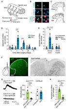
          Peripheral nerve lesions provoke apoptosis in the dorsal horn of the spinal cord. The cause of cell death, the involvement of neurons, and the relevance for the processing of somatosensory information are controversial. Here, we demonstrate in a mouse model of sciatic nerve injury that glutamate-induced neurodegeneration and loss of γ-aminobutyric acid (GABA)ergic interneurons in the superficial dorsal horn promote the transition from acute to chronic neuropathic pain. Conditional deletion of Grin1, the essential subunit of N-methyl-D-aspartate-type glutamate receptors (NMDARs), protects dorsal horn neurons from excitotoxicity and preserves GABAergic inhibition. Mice deficient in functional NMDARs exhibit normal nociceptive responses and acute pain after nerve injury, but this initial increase in pain sensitivity is reversible. Eliminating NMDARs fully prevents persistent pain-like behavior. Reduced pain in mice lacking proapoptotic Bax confirmed the significance of neurodegeneration. We conclude that NMDAR-mediated neuron death contributes to the development of chronic neuropathic pain.

          In Brief

          Dorsal horn neurons process somatosensory information, including pain. Inquimbert et al. utilized spatially restricted Grin1 knockout to show that NMDA-receptor-mediated excitatory input causes the degeneration of some dorsal horn neurons after nerve injury. Irreversible loss of GABAergic interneurons leads to a deficit in inhibition that promotes persistent pain hypersensitivity.

          Graphical Abstract

          Related collections

          Most cited references37

          • Record: found
          • Abstract: found
          • Article: not found

          Unbiased stereological estimation of the total number of neurons in thesubdivisions of the rat hippocampus using the optical fractionator.

          A stereological method for obtaining estimates of the total number of neurons in five major subdivisions of the rat hippocampus is described. The new method, the optical fractionator, combines two recent developments in stereology: a three-dimensional probe for counting neuronal nuclei, the optical disector, and a systematic uniform sampling scheme, the fractionator. The optical disector results in unbiased estimates of neuron number, i.e., estimates that are free of assumptions about neuron size and shape, are unaffected by lost caps and overprojection, and approach the true number of neurons in an unlimited manner as the number of samples is increased. The fractionator involves sampling a known fraction of a structural component. In the case of neuron number, a zero dimensional quantity, it provides estimates that are unaffected by shrinkage before, during, and after processing of the tissue. Because the fractionator involves systematic sampling, it also results in highly efficient estimates. Typically only 100-200 neurons must be counted in an animal to obtain a precision that is compatible with experimental studies. The methodology is compared with those used in earlier works involving estimates of neuron number in the rat hippocampus and a number of new stereological methods that have particular relevance to the quantitative study of the structure of the nervous system are briefly described in an appendix.
            Bookmark
            • Record: found
            • Abstract: found
            • Article: not found

            Spared nerve injury: an animal model of persistent peripheral neuropathic pain.

            Peripheral neuropathic pain is produced by multiple etiological factors that initiate a number of diverse mechanisms operating at different sites and at different times and expressed both within, and across different disease states. Unraveling the mechanisms involved requires laboratory animal models that replicate as far as possible, the different pathophysiological changes present in patients. It is unlikely that a single animal model will include the full range of neuropathic pain mechanisms. A feature of several animal models of peripheral neuropathic pain is partial denervation. In the most frequently used models a mixture of intact and injured fibers is created by loose ligation of either the whole (Bennett GJ, Xie YK. A peripheral mononeuropathy in rat that produces disorders of pain sensation like those seen in man. Pain 1988;33:87-107) or a tight ligation of a part (Seltzer Z, Dubner R, Shir Y. A novel behavioral model of neuropathic pain disorders produced in rats by partial sciatic nerve injury. Pain 1990;43:205-218) of a large peripheral nerve, or a tight ligation of an entire spinal segmental nerve (Kim SH, Chung JM. An experimental model for peripheral neuropathy produced by segmental spinal nerve ligation in the rat. Pain 1992;50:355-363). We have developed a variant of partial denervation, the spared nerve injury model. This involves a lesion of two of the three terminal branches of the sciatic nerve (tibial and common peroneal nerves) leaving the remaining sural nerve intact. The spared nerve injury model differs from the Chung spinal segmental nerve, the Bennett chronic constriction injury and the Seltzer partial sciatic nerve injury models in that the co-mingling of distal intact axons with degenerating axons is restricted, and it permits behavioral testing of the non-injured skin territories adjacent to the denervated areas. The spared nerve injury model results in early ( 6 months), robust (all animals are responders) behavioral modifications. The mechanical (von Frey and pinprick) sensitivity and thermal (hot and cold) responsiveness is increased in the ipsilateral sural and to a lesser extent saphenous territories, without any change in heat thermal thresholds. Crush injury of the tibial and common peroneal nerves produce similar early changes, which return, however to baseline at 7-9 weeks. The spared nerve injury model may provide, therefore, an additional resource for unraveling the mechanisms responsible for the production of neuropathic pain.
              Bookmark
              • Record: found
              • Abstract: found
              • Article: not found

              Green fluorescent protein expression and colocalization with calretinin, parvalbumin, and somatostatin in the GAD67-GFP knock-in mouse.

              Gamma-aminobutyric acid (GABA)ergic neurons in the central nervous system regulate the activity of other neurons and play a crucial role in information processing. To assist an advance in the research of GABAergic neurons, here we produced two lines of glutamic acid decarboxylase-green fluorescence protein (GAD67-GFP) knock-in mouse. The distribution pattern of GFP-positive somata was the same as that of the GAD67 in situ hybridization signal in the central nervous system. We encountered neither any apparent ectopic GFP expression in GAD67-negative cells nor any apparent lack of GFP expression in GAD67-positive neurons in the two GAD67-GFP knock-in mouse lines. The timing of GFP expression also paralleled that of GAD67 expression. Hence, we constructed a map of GFP distribution in the knock-in mouse brain. Moreover, we used the knock-in mice to investigate the colocalization of GFP with NeuN, calretinin (CR), parvalbumin (PV), and somatostatin (SS) in the frontal motor cortex. The proportion of GFP-positive cells among NeuN-positive cells (neocortical neurons) was approximately 19.5%. All the CR-, PV-, and SS-positive cells appeared positive for GFP. The CR-, PV, and SS-positive cells emitted GFP fluorescence at various intensities characteristics to them. The proportions of CR-, PV-, and SS-positive cells among GFP-positive cells were 13.9%, 40.1%, and 23.4%, respectively. Thus, the three subtypes of GABAergic neurons accounted for 77.4% of the GFP-positive cells. They accounted for 6.5% in layer I. In accord with unidentified GFP-positive cells, many medium-sized spherical somata emitting intense GFP fluorescence were observed in layer I. Copyright 2003 Wiley-Liss, Inc.
                Bookmark

                Author and article information

                Journal
                101573691
                39703
                Cell Rep
                Cell Rep
                Cell reports
                2211-1247
                23 November 2018
                29 May 2018
                29 May 2019
                : 23
                : 9
                : 2678-2689
                Affiliations
                [1 ]Centre National de la Recherche Scientifique, UPR 3212, Institut des Neurosciences Cellulaires et Intégratives and Université de Strasbourg, 67084 Strasbourg, France
                [2 ]F.M. Kirby Neurobiology Center, Boston Children’s Hospital and Department of Neurobiology, Harvard Medical School, Boston, MA 02115, USA
                [3 ]Departments of Anesthesiology and Pharmacology, Columbia University Medical Center, New York, NY 10032, USA
                [4 ]Institute of Pharmacology, Heidelberg University, 69120 Heidelberg, Germany
                [5 ]Department of Neurology and Neurosurgery, Johns Hopkins University School of Medicine, Baltimore, MD 21287, USA
                [6 ]Laboratory of Chromatin Biology and Epigenetics, Rockefeller University, New York, NY 10065, USA
                [7 ]Department of Structural and Functional Biology, State University of Campinas, Campinas, SP 13083-865, Brazil
                [8 ]Department of Genetic and Behavioral Neuroscience, Gunma University Graduate School of Medicine, Maebashi, Gunma 371-8511, Japan
                [9 ]Fishberg Department of Neuroscience and Friedman Brain Institute, Icahn School of Medicine at Mount Sinai, New York, NY 10029, USA
                [10 ]These authors contributed equally
                [11 ]Lead Contact
                Author notes

                AUTHOR CONTRIBUTIONS

                P.I., M.M., and J.S. designed the experiments. P.I., M.M., A.L., C.-K.T., J W., G.F.S., B.M.S., E.K., M.C.P.A., O.B., and N.G. collected and analyzed the results.Y.Y., C.D.A., and P.R.H. contributed to the study design and the interpretation of findings. P.I., M.M., and J.S. wrote the manuscript.

                Article
                NIHMS998205
                10.1016/j.celrep.2018.04.107
                6276118
                29847798
                ec811708-d40b-41b2-a7c1-f29e11f8d6a9

                This is an open access article under the CC BY-NC-ND license ( http://creativecommons.org/licenses/by-nc-nd/4.0/).

                History
                Categories
                Article

                Cell biology
                Cell biology

                Comments

                Comment on this article