13
views
0
recommends
+1 Recommend
0 collections
    0
    shares
      • Record: found
      • Abstract: found
      • Article: found
      Is Open Access

      HIF-2 α and Oct4 have synergistic effects on survival and myocardial repair of very small embryonic-like mesenchymal stem cells in infarcted hearts

      research-article

      Read this article at

      Bookmark
          There is no author summary for this article yet. Authors can add summaries to their articles on ScienceOpen to make them more accessible to a non-specialist audience.

          Abstract

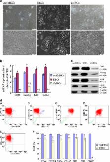
          Poor cell survival and limited functional benefits have restricted mesenchymal stem cell (MSC) efficacy for treating myocardial infarction (MI), suggesting that a better understanding of stem cell biology is needed. The transcription factor HIF-2 α is an essential regulator of the transcriptional response to hypoxia, which can interact with embryonic stem cells (ESCs) transcription factor Oct4 and modulate its signaling. Here, we obtained very small embryonic-like mesenchymal stem cells (vselMSCs) from MI patients, which possessed the very small embryonic-like stem cells' (VSELs) morphology as well as ESCs' pluripotency. Using microarray analysis, we compared HIF-2 α-regulated gene profiles in vselMSCs with ESC profiles and determined that HIF-2 α coexpressed Oct4 in vselMSCs similarly to ESCs. However, this coexpression was absent in unpurified MSCs (uMSCs). Under hypoxic condition, vselMSCs exhibited stronger survival, proliferation and differentiation than uMSCs. Transplantation of vselMSCs caused greater improvement in cardiac function and heart remodeling in the infarcted rats. We further demonstrated that HIF-2 α and Oct4 jointly regulate their relative downstream gene expressions, including Bcl2 and Survivin; the important pluripotent markers Nanog, Klf4, and Sox2; and Ang-1, bFGF, and VEGF, promoting angiogenesis and engraftment. Importantly, these effects were generally magnified by upregulation of HIF-2 α and Oct4 induced by HIF-2 α or Oct4 overexpression, and the greatest improvements were elicited after co-overexpressing HIF-2 α and Oct4; overexpressing one transcription factor while silencing the other canceled this increase, and HIF-2 α or Oct4 silencing abolished these effects. Together, these findings demonstrated that HIF-2 α in vselMSCs cooperated with Oct4 in survival and function. The identification of the cooperation between HIF-2 α and Oct4 will lead to deeper characterization of the downstream targets of this interaction in vselMSCs and will have novel pathophysiological implications for the repair of infarcted myocardium.

          Related collections

          Most cited references26

          • Record: found
          • Abstract: found
          • Article: not found

          Mesenchymal stem cells: biology, pathophysiology, translational findings, and therapeutic implications for cardiac disease.

          Mesenchymal stem cells (MSCs) are a prototypical adult stem cell with capacity for self-renewal and differentiation with a broad tissue distribution. Initially described in bone marrow, MSCs have the capacity to differentiate into mesoderm- and nonmesoderm-derived tissues. The endogenous role for MSCs is maintenance of stem cell niches (classically the hematopoietic), and as such, MSCs participate in organ homeostasis, wound healing, and successful aging. From a therapeutic perspective, and facilitated by the ease of preparation and immunologic privilege, MSCs are emerging as an extremely promising therapeutic agent for tissue regeneration. Studies in animal models of myocardial infarction have demonstrated the ability of transplanted MSCs to engraft and differentiate into cardiomyocytes and vasculature cells, recruit endogenous cardiac stem cells, and secrete a wide array of paracrine factors. Together, these properties can be harnessed to both prevent and reverse remodeling in the ischemically injured ventricle. In proof-of-concept and phase I clinical trials, MSC therapy improved left ventricular function, induced reverse remodeling, and decreased scar size. This article reviews the current understanding of MSC biology, mechanism of action in cardiac repair, translational findings, and early clinical trial data of MSC therapy for cardiac disease.
            Bookmark
            • Record: found
            • Abstract: found
            • Article: not found

            HIF-2alpha regulates Oct-4: effects of hypoxia on stem cell function, embryonic development, and tumor growth.

            The division, differentiation, and function of stem cells and multipotent progenitors are influenced by complex signals in the microenvironment, including oxygen availability. Using a genetic "knock-in" strategy, we demonstrate that targeted replacement of the oxygen-regulated transcription factor HIF-1alpha with HIF-2alpha results in expanded expression of HIF-2alpha-specific target genes including Oct-4, a transcription factor essential for maintaining stem cell pluripotency. We show that HIF-2alpha, but not HIF-1alpha, binds to the Oct-4 promoter and induces Oct-4 expression and transcriptional activity, thereby contributing to impaired development in homozygous Hif-2alpha KI/KI embryos, defective hematopoietic stem cell differentiation in embryoid bodies, and large embryonic stem cell (ES)-derived tumors characterized by altered cellular differentiation. Furthermore, loss of HIF-2alpha severely reduces the number of embryonic primordial germ cells, which require Oct-4 expression for survival and/or maintenance. These results identify Oct-4 as a HIF-2alpha-specific target gene and indicate that HIF-2alpha can regulate stem cell function and/or differentiation through activation of Oct-4, which in turn contributes to HIF-2alpha's tumor promoting activity.
              Bookmark
              • Record: found
              • Abstract: found
              • Article: not found

              A Phase II Dose-Escalation Study of Allogeneic Mesenchymal Precursor Cells in Patients With Ischemic or Nonischemic Heart Failure.

              Allogeneic mesenchymal precursor cells (MPCs) have been effective in large animal models of ischemic and nonischemic heart failure (HF).
                Bookmark

                Author and article information

                Journal
                Cell Death Dis
                Cell Death Dis
                Cell Death & Disease
                Nature Publishing Group
                2041-4889
                January 2017
                12 January 2017
                1 January 2017
                : 8
                : 1
                : e2548
                Affiliations
                [1 ]Department of Cardiology, the Third Affiliated Hospital of Southern Medical University , 183 West Zhongshan Road, Tianhe District, Guangzhou 510630, China
                [2 ]Department of Cardiology, Dahua Hospital , 901 Laohumin Rd, Xuhui District, Shanghai 200237, China
                [3 ]Department of Cardiology, Yangpu Hospital, Tongji Univercity School of Medicine , 450 Tengyue Rd, Shanghai 200090, China
                [4 ]Central Laboratory, Yangpu Hospital, Tongji Univercity School of Medicine , 450 Tengyue Rd, Shanghai 200090, China
                Author notes
                [* ]Department of Cardiology, the Third Affiliated Hospital of Southern Medical University , 183 West Zhongshan Road, Tianhe District, Guangzhou 510630, China. Tel: +86 1 20 62784342; Fax: +86 21 65696249; E-mail: shaohengzh67@ 123456163.com
                [* ]Central Laboratory, Yangpu Hospital, Tongji Univercity School of Medicine , 450 Tengyue Rd, Shanghai 200090, China. Tel: +86 21 65690520 281; Fax: +86 21 65673901; E-mail: xinpanpx@ 123456163.comm
                [5]

                These authors contributed equally to this work.

                Article
                cddis2016480
                10.1038/cddis.2016.480
                5386383
                28079892
                f34c5210-0fc3-48b6-9a80-ff19cbad9664
                Copyright © 2017 The Author(s)

                Cell Death and Disease is an open-access journal published by Nature Publishing Group. This work is licensed under a Creative Commons Attribution 4.0 International License. The images or other third party material in this article are included in the article's Creative Commons license, unless indicated otherwise in the credit line; if the material is not included under the Creative Commons license, users will need to obtain permission from the license holder to reproduce the material. To view a copy of this license, visit http://creativecommons.org/licenses/by/4.0/

                History
                : 05 September 2016
                : 08 December 2016
                : 13 December 2016
                Categories
                Original Article

                Cell biology
                Cell biology

                Comments

                Comment on this article