31
views
0
recommends
+1 Recommend
0 collections
    0
    shares
      • Record: found
      • Abstract: found
      • Article: found
      Is Open Access

      Seipin regulates ER–lipid droplet contacts and cargo delivery

      research-article

      Read this article at

      Bookmark
          There is no author summary for this article yet. Authors can add summaries to their articles on ScienceOpen to make them more accessible to a non-specialist audience.

          Abstract

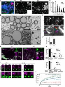
          Seipin is an endoplasmic reticulum ( ER) membrane protein implicated in lipid droplet ( LD) biogenesis and mutated in severe congenital lipodystrophy ( BSCL2). Here, we show that seipin is stably associated with nascent ERLD contacts in human cells, typically via one mobile focal point per LD. Seipin appears critical for such contacts since ERLD contacts were completely missing or morphologically aberrant in seipin knockout and BSCL2 patient cells. In parallel, LD mobility was increased and protein delivery from the ER to LDs to promote LD growth was decreased. Moreover, while growing LDs normally acquire lipid and protein constituents from the ER, this process was compromised in seipin‐deficient cells. In the absence of seipin, the initial synthesis of neutral lipids from exogenous fatty acid was normal, but fatty acid incorporation into neutral lipids in cells with pre‐existing LDs was impaired. Together, our data suggest that seipin helps to connect newly formed LDs to the ER and that by stabilizing ERLD contacts seipin facilitates the incorporation of protein and lipid cargo into growing LDs in human cells.

          Related collections

          Most cited references34

          • Record: found
          • Abstract: found
          • Article: not found

          Triacylglycerol synthesis enzymes mediate lipid droplet growth by relocalizing from the ER to lipid droplets.

          Lipid droplets (LDs) store metabolic energy and membrane lipid precursors. With excess metabolic energy, cells synthesize triacylglycerol (TG) and form LDs that grow dramatically. It is unclear how TG synthesis relates to LD formation and growth. Here, we identify two LD subpopulations: smaller LDs of relatively constant size, and LDs that grow larger. The latter population contains isoenzymes for each step of TG synthesis. Glycerol-3-phosphate acyltransferase 4 (GPAT4), which catalyzes the first and rate-limiting step, relocalizes from the endoplasmic reticulum (ER) to a subset of forming LDs, where it becomes stably associated. ER-to-LD targeting of GPAT4 and other LD-localized TG synthesis isozymes is required for LD growth. Key features of GPAT4 ER-to-LD targeting and function in LD growth are conserved between Drosophila and mammalian cells. Our results explain how TG synthesis is coupled with LD growth and identify two distinct LD subpopulations based on their capacity for localized TG synthesis. Copyright © 2013 Elsevier Inc. All rights reserved.
            Bookmark
            • Record: found
            • Abstract: found
            • Article: not found

            Engineered ascorbate peroxidase as a genetically-encoded reporter for electron microscopy

            Electron microscopy (EM) is the standard method for imaging cellular structures with nanometer resolution, but existing genetic tags are inactive in most cellular compartments 1 or require light and are difficult to use 2 . Here we report the development of a simple and robust EM genetic tag, called “APEX,” that is active in all cellular compartments and does not require light. APEX is a monomeric 28 kDa peroxidase that withstands strong EM fixation to give excellent ultrastructural preservation. We demonstrate the utility of APEX for high-resolution EM imaging of a variety of mammalian organelles and specific proteins. We also fused APEX to the N- or C-terminus of the mitochondrial calcium uniporter (MCU), a newly identified channel whose topology is disputed 3,4 . MCU-APEX and APEX-MCU give EM contrast exclusively in the mitochondrial matrix, suggesting that both the N-and C-termini of MCU face the matrix.
              Bookmark
              • Record: found
              • Abstract: found
              • Article: not found

              The lipodystrophy protein seipin is found at endoplasmic reticulum lipid droplet junctions and is important for droplet morphology.

              Lipodystrophy is a disorder characterized by a loss of adipose tissue often accompanied by severe hypertriglyceridemia, insulin resistance, diabetes, and fatty liver. It can be inherited or acquired. The most severe inherited form is Berardinelli-Seip Congenital Lipodystrophy Type 2, associated with mutations in the BSCL2 gene. BSCL2 encodes seipin, the function of which has been entirely unknown. We now report the identification of yeast BSCL2/seipin through a screen to detect genes important for lipid droplet morphology. The absence of yeast seipin results in irregular lipid droplets often clustered alongside proliferated endoplasmic reticulum (ER); giant lipid droplets are also seen. Many small irregular lipid droplets are also apparent in fibroblasts from a BSCL2 patient. Human seipin can functionally replace yeast seipin, but a missense mutation in human seipin that causes lipodystrophy, or corresponding mutations in the yeast gene, render them unable to complement. Yeast seipin is localized in the ER, where it forms puncta. Almost all lipid droplets appear to be on the ER, and seipin is found at these junctions. Therefore, we hypothesize that seipin is important for droplet maintenance and perhaps assembly. In addition to detecting seipin, the screen identified 58 other genes whose deletions cause aberrant lipid droplets, including 2 genes encoding proteins known to activate lipin, a lipodystrophy locus in mice, and 16 other genes that are involved in endosomal-lysosomal trafficking. The genes identified in our screen should be of value in understanding the pathway of lipid droplet biogenesis and maintenance and the cause of some lipodystrophies.
                Bookmark

                Author and article information

                Contributors
                elina.ikonen@helsinki.fi
                Journal
                EMBO J
                EMBO J
                10.1002/(ISSN)1460-2075
                EMBJ
                embojnl
                The EMBO Journal
                John Wiley and Sons Inc. (Hoboken )
                0261-4189
                1460-2075
                22 November 2016
                15 December 2016
                22 November 2016
                : 35
                : 24 ( doiID: 10.1002/embj.v35.24 )
                : 2699-2716
                Affiliations
                [ 1 ] Department of Anatomy Faculty of MedicineUniversity of Helsinki HelsinkiFinland
                [ 2 ]Minerva Foundation Institute for Medical Research HelsinkiFinland
                [ 3 ] Institute of BiotechnologyUniversity of Helsinki HelsinkiFinland
                [ 4 ] Sorbonne Universités UPMC Univ Paris 6 Inserm UMR_S938 Saint‐Antoine Research Center Institute of Cardiometabolism And NutritionAP‐HP, Saint‐Antoine Hospital Department of Molecular Biology and Genetics ParisFrance
                [ 5 ] l'Institut du ThoraxINSERM CNRS UNIV Nantes NantesFrance
                [ 6 ] LIMES InstituteBonn University BonnGermany
                Author notes
                [*] [* ]Corresponding author. Tel: +358 02941 25277; Fax: +358 02941 25261; E‐mail: elina.ikonen@ 123456helsinki.fi
                [†]

                These authors contributed equally to this work

                Author information
                http://orcid.org/0000-0003-1663-0704
                Article
                EMBJ201695170
                10.15252/embj.201695170
                5167346
                27879284
                fd73d51f-e39b-4fe2-929e-37bc3df8012f
                © 2016 The Authors. Published under the terms of the CC BY NC ND 4.0 license

                This is an open access article under the terms of the Creative Commons Attribution‐NonCommercial‐NoDerivs 4.0 License, which permits use and distribution in any medium, provided the original work is properly cited, the use is non‐commercial and no modifications or adaptations are made.

                History
                : 20 July 2016
                : 22 October 2016
                : 24 October 2016
                Page count
                Figures: 14, Tables: 0, Pages: 18, Words: 12252
                Funding
                Funded by: Academy of Finland
                Award ID: 272130
                Award ID: 282192
                Award ID: 284667
                Award ID: 131650
                Award ID: 1287975
                Funded by: The Finnish Medical Foundation
                Funded by: Helsinki Biomedical Graduate School
                Funded by: Sigrid Juselius Foundation
                Funded by: Biocenter Finland
                Funded by: Institut National de la Santé et de la Recherche Médicale
                Funded by: Aide aux Jeunes Diabétiques
                Funded by: Société Francophone du Diabète
                Funded by: Deutsche Forschungsgemeinschaft
                Award ID: SFB645
                Categories
                Article
                Articles
                Custom metadata
                2.0
                embj201695170
                15 December 2016
                Converter:WILEY_ML3GV2_TO_NLMPMC version:5.0.0 mode:remove_FC converted:19.12.2016

                Molecular biology
                electron microscopy,lipid storage,lipodystrophy,membrane contact sites,membrane & intracellular transport

                Comments

                Comment on this article