74
views
0
recommends
+1 Recommend
0 collections
    0
    shares
      • Record: found
      • Abstract: found
      • Article: found
      Is Open Access

      Cone-beam computed tomographic evaluation of styloid process: a retrospective study of 208 patients with orofacial pain

      research-article

      Read this article at

      Bookmark
          There is no author summary for this article yet. Authors can add summaries to their articles on ScienceOpen to make them more accessible to a non-specialist audience.

          Abstract

          Introduction

          The purpose of this study was to assess the structural characteristics of styloid process (SP) by cone-beam computed tomography (CBCT) examination in a patient population suffering from orofacial pain. The second aim was to assess the prevalence of elongated SP and its relation to gender, site and subjective symptoms in the study population.

          Methods

          Clinical and radiographic records of 208 patients were evaluated retrospectively. Radiological examinations including measurements of the structure, length, and medial angulations of SP were performed on CBCT images.

          Results

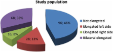
          Out of 208 patients, 96 (46%) had not-elongated SP, 28 (13%) had left side, 16 (8%) had right side, and 68 (33%) had bilateral elongation of SP. The patients with elongated SP had significantly decreased angle values. There were no statistically significant differences in length values of SP between males and females in both groups. Significantly increased prevalence of symptoms except headache was observed in patients with elongated SP.

          Conclusions

          This study presents the CBCT as an alternative method to CT or panoramic radiographs for the measurement and the assessment of the styloid process. Patients suffering from orofacial pain, who also had elongated SP, had increased rate of corresponding neurological complaints compared with non-elongated ones.

          Related collections

          Most cited references18

          • Record: found
          • Abstract: found
          • Article: not found

          Comparative dosimetry of dental CBCT devices and 64-slice CT for oral and maxillofacial radiology.

          This study compares 2 measures of effective dose, E(1990) and E(2007), for 8 dentoalveolar and maxillofacial cone-beam computerized tomography (CBCT) units and a 64-slice multidetector CT (MDCT) unit. Average tissue-absorbed dose, equivalent dose, and effective dose were calculated using thermoluminescent dosimeter chips in a radiation analog dosimetry phantom. Effective doses were derived using 1990 and the superseding 2007 International Commission on Radiological Protection (ICRP) recommendations. Large-field of view (FOV) CBCT E(2007) ranged from 68 to 1,073 microSv. Medium-FOV CBCT E(2007) ranged from 69 to 560 microSv, whereas a similar-FOV MDCT produced 860 microSv. The E(2007) calculations were 23% to 224% greater than E(1990). The 2007 recommendations of the ICRP, which include salivary glands, extrathoracic region, and oral mucosa in the calculation of effective dose, result in an upward reassessment of fatal cancer risk from oral and maxillofacial radiographic examinations. Dental CBCT can be recommended as a dose-sparing technique in comparison with alternative medical CT scans for common oral and maxillofacial radiographic imaging tasks.
            Bookmark
            • Record: found
            • Abstract: found
            • Article: not found

            Incidence of the type and calcification patterns in patients with elongated styloid process.

            We investigated the incidence of elongated styloid process (Eagle's syndrome) using panoramic radiographs taken of 860 patients referred to our clinic. Any styloid process identified was classified according to its length, type and the pattern of calcification. Fifty-nine elongated styloid processes were identified in 32 patients (3.7%), most being bilateral; 24 patients were female and eight were male (female/male ratio: 3:1). The mean age of these patients was 43 +/- 14 years (range: 18 - 78 years). Type I (elongated) was the most frequent type on both sides (42/59); and the most frequent patterns of calcification were partially calcified on the left side (18/59) and completely calcified on the right side (16/59). Only two patients were symptomatic. A corrected differential diagnosis is important to distinguish elongated styloid process from other pathologies with partially overlapping symptoms. We would recommend that clinicians consider the possibility of Eagle's syndrome when both the clinical and radiographic evidence support this diagnosis.
              Bookmark
              • Record: found
              • Abstract: found
              • Article: not found

              Elongated and mineralized stylohyoid ligament complex: a proposed classification and report of a case of Eagle's syndrome.

              A classification of the radiographic appearance of elongated and mineralized stylohyoid ligament complexes based on three types of complexes--Type I, elongated; Type II, pseudoarticulated; and Type III, segmented--is proposed. These types are further described by a pattern of calcification: calcified outline, partially calcified, nodular, and completely calcified. The classification is illustrated in a case of Eagle's syndrome in a 55-year-old Mexican-American man with symptoms of chronic otalgia and cephalgia. The surgical management and follow-up of this patient are discussed.
                Bookmark

                Author and article information

                Contributors
                Journal
                Head Face Med
                Head Face Med
                Head & Face Medicine
                BioMed Central
                1746-160X
                2014
                15 February 2014
                : 10
                : 5
                Affiliations
                [1 ]Department of Oral and Maxillofacial Radiology, Faculty of Dentistry, Çukurova University, Saricam, 01330 Adana, Turkey
                [2 ]Department of Oral and Maxillofacial Surgery, Faculty of Dentistry, Çukurova University, Adana, Turkey
                [3 ]Department of Neurology, Faculty of Medicine, Çukurova University, Adana, Turkey
                Article
                1746-160X-10-5
                10.1186/1746-160X-10-5
                3943457
                24528515
                fd8af101-c198-4a22-a60f-3440aead0e22
                Copyright © 2014 Öztunç et al.; licensee BioMed Central Ltd.

                This is an Open Access article distributed under the terms of the Creative Commons Attribution License ( http://creativecommons.org/licenses/by/2.0), which permits unrestricted use, distribution, and reproduction in any medium, provided the original work is properly credited. The Creative Commons Public Domain Dedication waiver ( http://creativecommons.org/publicdomain/zero/1.0/) applies to the data made available in this article, unless otherwise stated.

                History
                : 3 December 2013
                : 13 February 2014
                Categories
                Research

                Orthopedics
                cone-beam computed tomography,eagle’s syndrome,elongated styloid process,orofacial pain,retrospective study

                Comments

                Comment on this article