9
views
0
recommends
+1 Recommend
0 collections
    0
    shares
      • Record: found
      • Abstract: found
      • Article: found
      Is Open Access

      The temporal dynamics of chromosome instability in ovarian cancer cell lines and primary patient samples

      research-article

      Read this article at

      Bookmark
          There is no author summary for this article yet. Authors can add summaries to their articles on ScienceOpen to make them more accessible to a non-specialist audience.

          Abstract

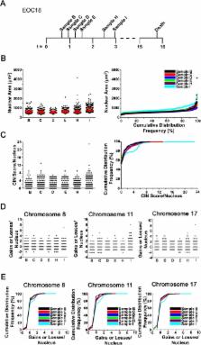
          Epithelial ovarian cancer (EOC) is the most prevalent form of ovarian cancer and has the highest mortality rate. Novel insight into EOC is required to minimize the morbidity and mortality rates caused by recurrent, drug resistant disease. Although numerous studies have evaluated genome instability in EOC, none have addressed the putative role chromosome instability (CIN) has in disease progression and drug resistance. CIN is defined as an increase in the rate at which whole chromosomes or large parts thereof are gained or lost, and can only be evaluated using approaches capable of characterizing genetic or chromosomal heterogeneity within populations of cells. Although CIN is associated with numerous cancer types, its prevalence and dynamics in EOC is unknown. In this study, we assessed CIN within serial samples collected from the ascites of five EOC patients, and in two well-established ovarian cancer cell models of drug resistance (PEO1/4 and A2780s/cp). We quantified and compared CIN (as measured by nuclear areas and CIN Score (CS) values) within and between serial samples to glean insight into the association and dynamics of CIN within EOC, with a particular focus on resistant and recurrent disease. Using quantitative, single cell analyses we determined that CIN is associated with every sample evaluated and further show that many EOC samples exhibit a large degree of nuclear size and CS value heterogeneity. We also show that CIN is dynamic and generally increases within resistant disease. Finally, we show that both drug resistance models (PEO1/4 and A2780s/cp) exhibit heterogeneity, albeit to a much lesser extent. Surprisingly, the two cell line models exhibit remarkably similar levels of CIN, as the nuclear areas and CS values are largely overlapping between the corresponding paired lines. Accordingly, these data suggest CIN may represent a novel biomarker capable of monitoring changes in EOC progression associated with drug resistance.

          Author summary

          Ovarian cancer is one of the most lethal cancers in women due to the high prevalence of drug resistant disease. New insight into the biology causing drug resistance is required to reduce death rates associated with the disease. In many cancer types, chromosome instability (CIN; or abnormal numbers of chromosomes) is associated with aggressive tumours, the acquisition of multi-drug resistance and poor patient outcome, yet CIN is poorly characterized in ovarian cancer. Here, we employ a new microscopy-based approach to examine the presence and dynamics of CIN within single cells obtained from ovarian cancer patients collected over time. We show that CIN is associated with every sample and further show that increases in CIN are associated with treatment and/or the development of drug (platinum) resistant disease. Although additional studies are required, these findings suggest CIN may be a new biomarker that can monitor disease progression, particularly in response to therapy. Finally, we demonstrate that CIN is low in two cellular models of drug resistant disease. Collectively, our findings strengthen the need for studies employing patient-derived samples to accurately assess the abnormal biology associated with the disease, and cautions the use of these cell models in studies related to drug resistance that may arise from CIN.

          Related collections

          Most cited references41

          • Record: found
          • Abstract: found
          • Article: not found

          Genetic instabilities in human cancers.

          Whether and how human tumours are genetically unstable has been debated for decades. There is now evidence that most cancers may indeed be genetically unstable, but that the instability exists at two distinct levels. In a small subset of tumours, the instability is observed at the nucleotide level and results in base substitutions or deletions or insertions of a few nucleotides. In most other cancers, the instability is observed at the chromosome level, resulting in losses and gains of whole chromosomes or large portions thereof. Recognition and comparison of these instabilities are leading to new insights into tumour pathogenesis.
            Bookmark
            • Record: found
            • Abstract: found
            • Article: not found

            Mechanisms of chromosomal instability.

            Most solid tumors are aneuploid, having a chromosome number that is not a multiple of the haploid number, and many frequently mis-segregate whole chromosomes in a phenomenon called chromosomal instability (CIN). CIN positively correlates with poor patient prognosis, indicating that reduced mitotic fidelity contributes to cancer progression by increasing genetic diversity among tumor cells. Here, we review the mechanisms underlying CIN, which include defects in chromosome cohesion, mitotic checkpoint function, centrosome copy number, kinetochore-microtubule attachment dynamics, and cell-cycle regulation. Understanding these mechanisms provides insight into the cellular consequences of CIN and reveals the possibility of exploiting CIN in cancer therapy. 2010 Elsevier Ltd. All rights reserved.
              Bookmark
              • Record: found
              • Abstract: found
              • Article: found
              Is Open Access

              Getting to Know Ovarian Cancer Ascites: Opportunities for Targeted Therapy-Based Translational Research

              More than one third of ovarian cancer patients present with ascites at diagnosis, and almost all have ascites at recurrence. The presence of ascites correlates with the peritoneal spread of ovarian cancer and is associated with poor disease prognosis. Malignant ascites acts as a reservoir of a complex mixture of soluble factors and cellular components which provide a pro-inflammatory and tumor-promoting microenvironment for the tumor cells. Subpopulations of these tumor cells exhibit cancer stem-like phenotypes, possess enhanced resistance to therapies and the capacity for distal metastatic spread and recurrent disease. Thus, ascites-derived malignant cells and the ascites microenvironment represent a major source of morbidity and mortality for ovarian cancer patients. This review focuses on recent advances in our understanding of the molecular, cellular, and functional characteristics of the cellular populations within ascites and discusses their contributions to ovarian cancer metastasis, chemoresistance, and recurrence. We highlight in particular recent translational findings which have used primary ascites-derived tumor cells as a tool to understand the pathogenesis of the disease, yielding new insights and targets for therapeutic manipulation.
                Bookmark

                Author and article information

                Contributors
                Role: Editor
                Journal
                PLoS Genet
                PLoS Genet
                plos
                plosgen
                PLoS Genetics
                Public Library of Science (San Francisco, CA USA )
                1553-7390
                1553-7404
                4 April 2017
                April 2017
                : 13
                : 4
                : e1006707
                Affiliations
                [1 ]Department of Biochemistry and Medical Genetics, University of Manitoba, Winnipeg, Manitoba, Canada
                [2 ]Research Institute in Oncology and Hematology, CancerCare Manitoba, Winnipeg, Manitoba, Canada
                [3 ]Department of Obstetrics, Gynecology and Reproductive Sciences, University of Manitoba, Winnipeg, Manitoba, Canada
                Cleveland Clinic Genomic Medicine Institute, UNITED STATES
                Author notes

                The authors have declared that no competing interests exist.

                • Conceptualization: MWN KJM.

                • Formal analysis: SPG MN MWN KJM.

                • Investigation: SPG MN ZL JLA.

                • Resources: ADA.

                • Writing – original draft: SPG MWN KJM.

                • Writing – review & editing: SPG ADA MWN KJM.

                Author information
                http://orcid.org/0000-0002-0280-2555
                http://orcid.org/0000-0003-3291-8628
                Article
                PGENETICS-D-16-02398
                10.1371/journal.pgen.1006707
                5395197
                28376088
                0ff8489a-d2de-46b8-8f6a-af3c6659b53d
                © 2017 Penner-Goeke et al

                This is an open access article distributed under the terms of the Creative Commons Attribution License, which permits unrestricted use, distribution, and reproduction in any medium, provided the original author and source are credited.

                History
                : 26 October 2016
                : 20 March 2017
                Page count
                Figures: 8, Tables: 0, Pages: 24
                Funding
                Funded by: funder-id http://dx.doi.org/10.13039/501100000024, Canadian Institutes of Health Research;
                Award ID: MOP-115179
                Award Recipient :
                Funded by: funder-id http://dx.doi.org/10.13039/100008794, Research Manitoba;
                Award Recipient :
                Funded by: University of Manitoba
                Award ID: University Collaborative Research Grant
                Award Recipient :
                Funded by: funder-id http://dx.doi.org/10.13039/100009395, CancerCare Manitoba Foundation;
                Award ID: Summer Studentship
                Award Recipient :
                Funded by: funder-id http://dx.doi.org/10.13039/501100000038, Natural Sciences and Engineering Research Council of Canada;
                Award ID: Undergraduate Student Research Award
                Award Recipient :
                Funded by: funder-id http://dx.doi.org/10.13039/501100000038, Natural Sciences and Engineering Research Council of Canada;
                Award ID: RGPIN/401851
                Award Recipient :
                This work was funded by operating grants from the Canadian Institutes of Health Research (KJM; MOP-115179), the Natural Sciences and Engineering Research Council of Canada (KJM; RGPIN/401851), a Research Manitoba Operating Grant (MWN), University of Manitoba - University Collaborative Research Grant (MWN and KJM), a CancerCare Manitoba Foundation Summer Studentship (SPG) and a Natural Sciences and Engineering Research Council of Canada Undergraduate Student Research Award (MN). The funders had no role in study design, data collection and analysis, decision to publish, or preparation of the manuscript.
                Categories
                Research Article
                Medicine and Health Sciences
                Clinical Genetics
                Chromosomal Disorders
                Physical Sciences
                Mathematics
                Probability Theory
                Statistical Distributions
                Biology and Life Sciences
                Cell Biology
                Chromosome Biology
                Chromosomes
                Physical Sciences
                Chemistry
                Chemical Elements
                Platinum
                Medicine and Health Sciences
                Oncology
                Cancers and Neoplasms
                Gynecological Tumors
                Ovarian Cancer
                Biology and Life Sciences
                Genetics
                Gene Identification and Analysis
                Genetic Networks
                Computer and Information Sciences
                Network Analysis
                Genetic Networks
                Biology and Life Sciences
                Cell Biology
                Chromosome Biology
                Chromosomes
                Chromosome Structure and Function
                Biology and Life Sciences
                Cell Biology
                Chromosome Biology
                Chromosomes
                Chromosome Pairs
                Chromosome 8
                Custom metadata
                vor-update-to-uncorrected-proof
                2017-04-18
                All relevant data are within the paper and its Supporting Information files.

                Genetics
                Genetics

                Comments

                Comment on this article