2
views
0
recommends
+1 Recommend
0 collections
    0
    shares
      • Record: found
      • Abstract: found
      • Article: found
      Is Open Access

      YTHDC1-mediated augmentation of miR-30d in repressing pancreatic tumorigenesis via attenuation of RUNX1-induced transcriptional activation of Warburg effect

      research-article

      Read this article at

      Bookmark
          There is no author summary for this article yet. Authors can add summaries to their articles on ScienceOpen to make them more accessible to a non-specialist audience.

          Abstract

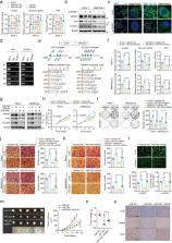
          Pancreatic ductal adenocarcinoma (PDAC) is one of the most lethal human cancers. It thrives in a malnourished environment; however, little is known about the mechanisms by which PDAC cells actively promote aerobic glycolysis to maintain their metabolic needs. Gene Expression Omnibus (GEO) was used to identify differentially expressed miRNAs. The expression pattern of miR-30d in normal and PDAC tissues was studied by in situ hybridization. The role of miR-30d/RUNX1 in vitro and in vivo was evaluated by CCK8 assay and clonogenic formation as well as transwell experiment, subcutaneous xenograft model and liver metastasis model, respectively. Glucose uptake, ATP and lactate production were tested to study the regulatory effect of miR-30d/RUNX1 on aerobic glycolysis in PDAC cells. Quantitative real-time PCR, western blot, Chip assay, promoter luciferase activity, RIP, MeRIP, and RNA stability assay were used to explore the molecular mechanism of YTHDC1/miR-30d/RUNX1 in PDAC. Here, we discover that miR-30d expression was remarkably decreased in PDAC tissues and associated with good prognosis, contributed to the suppression of tumor growth and metastasis, and attenuation of Warburg effect. Mechanistically, the m 6A reader YTHDC1 facilitated the biogenesis of mature miR-30d via m 6A-mediated regulation of mRNA stability. Then, miR-30d inhibited aerobic glycolysis through regulating SLC2A1 and HK1 expression by directly targeting the transcription factor RUNX1, which bound to the promoters of the SLC2A1 and HK1 genes. Moreover, miR-30d was clinically inversely correlated with RUNX1, SLC2A1 and HK1, which function as adverse prognosis factors for overall survival in PDAC tissues. Overall, we demonstrated that miR-30d is a functional and clinical tumor-suppressive gene in PDAC. Our findings further uncover that miR-30d is a novel target for YTHDC1 through m 6A modification, and miR-30d represses pancreatic tumorigenesis via suppressing aerobic glycolysis.

          Related collections

          Most cited references42

          • Record: found
          • Abstract: found
          • Article: not found

          m6A-dependent regulation of messenger RNA stability

          N6 -methyladenosine (m6A) is the most prevalent internal (non-cap) modification present in the messenger RNA (mRNA) of all higher eukaryotes 1,2 . Although essential to cell viability and development 3–5 , the exact role of m6A modification remains to be determined. The recent discovery of two m6A demethylases in mammalian cells highlighted the importance of m6A in basic biological functions and disease 6–8 . Here we show that m6A is selectively recognized by the human YTH domain family 2 (YTHDF2) protein to regulate mRNA degradation. We identified over 3,000 cellular RNA targets of YTHDF2, most of which are mRNAs, but which also include non-coding RNAs, with a conserved core motif of G(m6A)C. We further establish the role of YTHDF2 in RNA metabolism, showing that binding of YTHDF2 results in the localization of bound mRNA from the translatable pool to mRNA decay sites, such as processing bodies 9 . The C-terminal domain of YTHDF2 selectively binds to m6A-containing mRNA whereas the N-terminal domain is responsible for the localization of the YTHDF2-mRNA complex to cellular RNA decay sites. Our results indicate that the dynamic m6A modification is recognized by selective-binding proteins to affect the translation status and lifetime of mRNA.
            Bookmark
            • Record: found
            • Abstract: found
            • Article: not found

            Reading, writing and erasing mRNA methylation

            RNA methylation to form N6-methyladenosine (m6A) in mRNA accounts for the most abundant mRNA internal modification and has emerged as a widespread regulatory mechanism that controls gene expression in diverse physiological processes. Transcriptome-wide m6A mapping has revealed the distribution and pattern of m6A in cellular RNAs, referred to as the epitranscriptome. These maps have revealed the specific mRNAs that are regulated by m6A, providing mechanistic links connecting m6A to cellular differentiation, cancer progression and other processes. The effects of m6A on mRNA are mediated by an expanding list of m6A readers and m6A writer-complex components, as well as potential erasers that currently have unclear relevance to m6A prevalence in the transcriptome. Here we review new and emerging methods to characterize and quantify the epitranscriptome, and we discuss new concepts - in some cases, controversies - regarding our understanding of the mechanisms and functions of m6A readers, writers and erasers.
              Bookmark
              • Record: found
              • Abstract: found
              • Article: found

              Oncogenic Kras Maintains Pancreatic Tumors through Regulation of Anabolic Glucose Metabolism

              Tumor maintenance relies on continued activity of driver oncogenes, although their rate-limiting role is highly context dependent. Oncogenic Kras mutation is the signature event in pancreatic ductal adenocarcinoma (PDAC), serving a critical role in tumor initiation. Here, an inducible Kras(G12D)-driven PDAC mouse model establishes that advanced PDAC remains strictly dependent on Kras(G12D) expression. Transcriptome and metabolomic analyses indicate that Kras(G12D) serves a vital role in controlling tumor metabolism through stimulation of glucose uptake and channeling of glucose intermediates into the hexosamine biosynthesis and pentose phosphate pathways (PPP). These studies also reveal that oncogenic Kras promotes ribose biogenesis. Unlike canonical models, we demonstrate that Kras(G12D) drives glycolysis intermediates into the nonoxidative PPP, thereby decoupling ribose biogenesis from NADP/NADPH-mediated redox control. Together, this work provides in vivo mechanistic insights into how oncogenic Kras promotes metabolic reprogramming in native tumors and illuminates potential metabolic targets that can be exploited for therapeutic benefit in PDAC. Copyright © 2012 Elsevier Inc. All rights reserved.
                Bookmark

                Author and article information

                Contributors
                lilei2968900@163.com
                meng_xiangjun@yahoo.com
                Journal
                Cell Death Differ
                Cell Death Differ
                Cell Death and Differentiation
                Nature Publishing Group UK (London )
                1350-9047
                1476-5403
                21 May 2021
                21 May 2021
                November 2021
                : 28
                : 11
                : 3105-3124
                Affiliations
                [1 ]GRID grid.16821.3c, ISNI 0000 0004 0368 8293, Department of Gastroenterology, Shanghai Nineth People’s Hospital, School of Medicine, , Shanghai Jiao Tong University, ; Shanghai, China
                [2 ]GRID grid.16821.3c, ISNI 0000 0004 0368 8293, Digestive Disease Research and Clinical Translation Center, , Shanghai Jiaotong University, ; Shanghai, China
                [3 ]GRID grid.16821.3c, ISNI 0000 0004 0368 8293, Division of Gastroenterology and Hepatology, Key Laboratory Gastroenterology and Hepatology, Ministry of Health, State Key Laboratory for Oncogenes and Related Genes, Renji Hospital, School of Medicine, , Shanghai Jiao Tong University, Shanghai Institute of Digestive Disease, ; Shanghai, China
                Article
                804
                10.1038/s41418-021-00804-0
                8563797
                34021267
                15e9feeb-39f6-4a1e-a7f6-f8f137f5cbbc
                © The Author(s) 2021

                Open Access This article is licensed under a Creative Commons Attribution 4.0 International License, which permits use, sharing, adaptation, distribution and reproduction in any medium or format, as long as you give appropriate credit to the original author(s) and the source, provide a link to the Creative Commons license, and indicate if changes were made. The images or other third party material in this article are included in the article’s Creative Commons license, unless indicated otherwise in a credit line to the material. If material is not included in the article’s Creative Commons license and your intended use is not permitted by statutory regulation or exceeds the permitted use, you will need to obtain permission directly from the copyright holder. To view a copy of this license, visit http://creativecommons.org/licenses/by/4.0/.

                History
                : 17 September 2020
                : 1 May 2021
                : 5 May 2021
                Funding
                Funded by: FundRef https://doi.org/10.13039/501100001809, National Natural Science Foundation of China (National Science Foundation of China);
                Award ID: NO. 81802304
                Award Recipient :
                Categories
                Article
                Custom metadata
                © ADMC Associazione Differenziamento e Morte Cellulare 2021

                Cell biology
                oncogenes,cancer metabolism
                Cell biology
                oncogenes, cancer metabolism

                Comments

                Comment on this article