Inviting an author to review:
Find an author and click ‘Invite to review selected article’ near their name.
Search for authorsSearch for similar articles
12
views
0
recommends
+1 Recommend
0 collections
    0
    shares
      • Record: found
      • Abstract: found
      • Article: found
      Is Open Access

      Modeling radiation injury-induced cell death and countermeasure drug responses in a human Gut-on-a-Chip

      research-article

      Read this article at
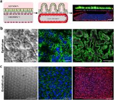
      Bookmark
          There is no author summary for this article yet. Authors can add summaries to their articles on ScienceOpen to make them more accessible to a non-specialist audience.

          Abstract

          Studies on human intestinal injury induced by acute exposure to γ-radiation commonly rely on use of animal models because culture systems do not faithfully mimic human intestinal physiology. Here we used a human Gut-on-a-Chip (Gut Chip) microfluidic device lined by human intestinal epithelial cells and vascular endothelial cells to model radiation injury and assess the efficacy of radiation countermeasure drugs in vitro. Exposure of the Gut Chip to γ-radiation resulted in increased generation of reactive oxygen species, cytotoxicity, apoptosis, and DNA fragmentation, as well as villus blunting, disruption of tight junctions, and compromise of intestinal barrier integrity. In contrast, pre-treatment with a potential prophylactic radiation countermeasure drug, dimethyloxaloylglycine (DMOG), significantly suppressed all of these injury responses. Thus, the human Gut Chip may serve as an in vitro platform for studying radiation-induced cell death and associate gastrointestinal acute syndrome, in addition to screening of novel radio-protective medical countermeasure drugs.

          Related collections

          Most cited references57

          • Record: found
          • Abstract: found
          • Article: not found

          Intestinal Goblet Cells and Mucins in Health and Disease: Recent Insights and Progress

          The mucus layer coating the gastrointestinal tract is the front line of innate host defense, largely because of the secretory products of intestinal goblet cells. Goblet cells synthesize secretory mucin glycoproteins (MUC2) and bioactive molecules such as epithelial membrane-bound mucins (MUC1, MUC3, MUC17), trefoil factor peptides (TFF), resistin-like molecule β (RELMβ), and Fc-γ binding protein (Fcgbp). The MUC2 mucin protein forms trimers by disulfide bonding in cysteine-rich amino terminal von Willebrand factor (vWF) domains, coupled with crosslinking provided by TFF and Fcgbp proteins with MUC2 vWF domains, resulting in a highly viscous extracellular layer. Colonization by commensal intestinal microbiota is limited to an outer “loose” mucus layer, and interacts with the diverse oligosaccharides of mucin glycoproteins, whereas an “inner” adherent mucus layer is largely devoid of bacteria. Defective mucus layers resulting from lack of MUC2 mucin, mutated Muc2 mucin vWF domains, or from deletion of core mucin glycosyltransferase enzymes in mice result in increased bacterial adhesion to the surface epithelium, increased intestinal permeability, and enhanced susceptibility to colitis caused by dextran sodium sulfate. Changes in mucin gene expression and mucin glycan structures occur in cancers of the intestine, contributing to diverse biologic properties involved in the development and progression of cancer. Further research is needed on identification and functional significance of various components of mucus layers and the complex interactions among mucus layers, microbiota, epithelial cells, and the underlying innate and adaptive immunity. Further elucidation of the regulatory mechanisms involved in mucin changes in cancer and inflammation may lead to the development of novel therapeutic approaches.
            Bookmark
            • Record: found
            • Abstract: found
            • Article: not found

            Tumor response to radiotherapy regulated by endothelial cell apoptosis.

            About 50% of cancer patients receive radiation therapy. Here we investigated the hypothesis that tumor response to radiation is determined not only by tumor cell phenotype but also by microvascular sensitivity. MCA/129 fibrosarcomas and B16F1 melanomas grown in apoptosis-resistant acid sphingomyelinase (asmase)-deficient or Bax-deficient mice displayed markedly reduced baseline microvascular endothelial apoptosis and grew 200 to 400% faster than tumors on wild-type microvasculature. Thus, endothelial apoptosis is a homeostatic factor regulating angiogenesis-dependent tumor growth. Moreover, these tumors exhibited reduced endothelial apoptosis upon irradiation and, unlike tumors in wild-type mice, they were resistant to single-dose radiation up to 20 grays (Gy). These studies indicate that microvascular damage regulates tumor cell response to radiation at the clinically relevant dose range.
              Bookmark
              • Record: found
              • Abstract: found
              • Article: not found

              Endothelial apoptosis as the primary lesion initiating intestinal radiation damage in mice.

              Gastrointestinal (GI) tract damage by chemotherapy or radiation limits their efficacy in cancer treatment. Radiation has been postulated to target epithelial stem cells within the crypts of Lieberkühn to initiate the lethal GI syndrome. Here, we show in mouse models that microvascular endothelial apoptosis is the primary lesion leading to stem cell dysfunction. Radiation-induced crypt damage, organ failure, and death from the GI syndrome were prevented when endothelial apoptosis was inhibited pharmacologically by intravenous basic fibroblast growth factor (bFGF) or genetically by deletion of the acid sphingomyelinase gene. Endothelial, but not crypt, cells express FGF receptor transcripts, suggesting that the endothelial lesion occurs before crypt stem cell damage in the evolution of the GI syndrome. This study provides a basis for new approaches to prevent radiation damage to the bowel.
                Bookmark

                Author and article information

                Contributors
                don.ingber@wyss.harvard.edu
                Journal
                Cell Death Dis
                Cell Death Dis
                Cell Death & Disease
                Nature Publishing Group UK (London )
                2041-4889
                14 February 2018
                14 February 2018
                February 2018
                : 9
                : 2
                : 223
                Affiliations
                [1 ]ISNI 000000041936754X, GRID grid.38142.3c, Wyss Institute for Biologically Inspired Engineering at Harvard University, ; Boston, MA 02115 USA
                [2 ]ISNI 0000 0001 2181 4263, GRID grid.9983.b, Department of Bioengineering and iBB - Institute for Bioengineering and Biosciences, Instituto Superior Técnico, , Universidade de Lisboa, ; Lisboa, Portugal
                [3 ]ISNI 0000 0004 0378 8438, GRID grid.2515.3, Vascular Biology Program and Department of Surgery, , Boston Children’s Hospital and Harvard Medical School, ; Boston, MA 02115 USA
                [4 ]ISNI 0000 0004 1936 9924, GRID grid.89336.37, Department of Biomedical Engineering, , The University of Texas at Austin, ; Austin, TX 78712 USA
                [5 ]ISNI 000000041936754X, GRID grid.38142.3c, Harvard John A. Paulson School of Engineering and Applied Sciences, ; Cambridge, MA 02139 USA
                Author information
                http://orcid.org/0000-0002-5004-893X
                Article
                304
                10.1038/s41419-018-0304-8
                5833800
                29445080
                1b4880a9-3d45-474a-b198-94e3389c3ef0
                © The Author(s) 2018

                Open Access This article is licensed under a Creative Commons Attribution 4.0 International License, which permits use, sharing, adaptation, distribution and reproduction in any medium or format, as long as you give appropriate credit to the original author(s) and the source, provide a link to the Creative Commons license, and indicate if changes were made. The images or other third party material in this article are included in the article’s Creative Commons license, unless indicated otherwise in a credit line to the material. If material is not included in the article’s Creative Commons license and your intended use is not permitted by statutory regulation or exceeds the permitted use, you will need to obtain permission directly from the copyright holder. To view a copy of this license, visit http://creativecommons.org/licenses/by/4.0/.

                History
                : 3 August 2017
                : 13 December 2017
                : 12 January 2018
                Categories
                Article
                Custom metadata
                © The Author(s) 2018

                Cell biology
                Cell biology

                Comments

                Comment on this article