3
views
0
recommends
+1 Recommend
0 collections
    0
    shares
      • Record: found
      • Abstract: found
      • Article: found
      Is Open Access

      The molecular landscape of neural differentiation in the developing Drosophila brain revealed by targeted scRNA-seq and multi-informatic analysis

      research-article

      Read this article at
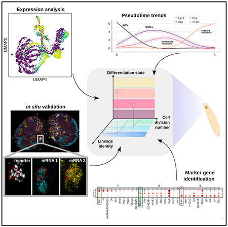
      Bookmark
          There is no author summary for this article yet. Authors can add summaries to their articles on ScienceOpen to make them more accessible to a non-specialist audience.

          SUMMARY

          The Drosophila type II neuroblast lineages present an attractive model to investigate the neurogenesis and differentiation process as they adapt to a process similar to that in the human outer subventricular zone. We perform targeted single-cell mRNA sequencing in third instar larval brains to study this process of the type II NB lineage. Combining prior knowledge, in silico analyses, and in situ validation, our multi-informatic investigation describes the molecular landscape from a single developmental snapshot. 17 markers are identified to differentiate distinct maturation stages. 30 markers are identified to specify the stem cell origin and/or cell division numbers of INPs, and at least 12 neuronal subtypes are identified. To foster future discoveries, we provide annotated tables of pairwise gene-gene correlation in single cells and MiCV, a web tool for interactively analyzing scRNA-seq datasets. Taken together, these resources advance our understanding of the neural differentiation process at the molecular level.

          In brief

          Using a combination of targeted scRNA-seq, in situ RNA staining, and a multi-informatic analysis paradigm, Michki et al. characterize the transcriptome landscape of thousands of type II neurons and their progenitors in the developing larval fruit fly brain.

          Graphical Abstract

          Related collections

          Most cited references81

          • Record: found
          • Abstract: found
          • Article: not found

          Fiji: an open-source platform for biological-image analysis.

          Fiji is a distribution of the popular open-source software ImageJ focused on biological-image analysis. Fiji uses modern software engineering practices to combine powerful software libraries with a broad range of scripting languages to enable rapid prototyping of image-processing algorithms. Fiji facilitates the transformation of new algorithms into ImageJ plugins that can be shared with end users through an integrated update system. We propose Fiji as a platform for productive collaboration between computer science and biology research communities.
            Bookmark
            • Record: found
            • Abstract: found
            • Article: not found

            STAR: ultrafast universal RNA-seq aligner.

            Accurate alignment of high-throughput RNA-seq data is a challenging and yet unsolved problem because of the non-contiguous transcript structure, relatively short read lengths and constantly increasing throughput of the sequencing technologies. Currently available RNA-seq aligners suffer from high mapping error rates, low mapping speed, read length limitation and mapping biases. To align our large (>80 billon reads) ENCODE Transcriptome RNA-seq dataset, we developed the Spliced Transcripts Alignment to a Reference (STAR) software based on a previously undescribed RNA-seq alignment algorithm that uses sequential maximum mappable seed search in uncompressed suffix arrays followed by seed clustering and stitching procedure. STAR outperforms other aligners by a factor of >50 in mapping speed, aligning to the human genome 550 million 2 × 76 bp paired-end reads per hour on a modest 12-core server, while at the same time improving alignment sensitivity and precision. In addition to unbiased de novo detection of canonical junctions, STAR can discover non-canonical splices and chimeric (fusion) transcripts, and is also capable of mapping full-length RNA sequences. Using Roche 454 sequencing of reverse transcription polymerase chain reaction amplicons, we experimentally validated 1960 novel intergenic splice junctions with an 80-90% success rate, corroborating the high precision of the STAR mapping strategy. STAR is implemented as a standalone C++ code. STAR is free open source software distributed under GPLv3 license and can be downloaded from http://code.google.com/p/rna-star/.
              Bookmark
              • Record: found
              • Abstract: found
              • Article: not found

              Systematic and integrative analysis of large gene lists using DAVID bioinformatics resources.

              DAVID bioinformatics resources consists of an integrated biological knowledgebase and analytic tools aimed at systematically extracting biological meaning from large gene/protein lists. This protocol explains how to use DAVID, a high-throughput and integrated data-mining environment, to analyze gene lists derived from high-throughput genomic experiments. The procedure first requires uploading a gene list containing any number of common gene identifiers followed by analysis using one or more text and pathway-mining tools such as gene functional classification, functional annotation chart or clustering and functional annotation table. By following this protocol, investigators are able to gain an in-depth understanding of the biological themes in lists of genes that are enriched in genome-scale studies.
                Bookmark

                Author and article information

                Journal
                101573691
                39703
                Cell Rep
                Cell Rep
                Cell reports
                2211-1247
                5 May 2021
                27 April 2021
                21 May 2021
                : 35
                : 4
                : 109039
                Affiliations
                [1 ]Biophysics LS&A, University of Michigan, Ann Arbor, MI, USA
                [2 ]Department of Cell and Developmental Biology, University of Michigan Medical School, Ann Arbor, MI, USA
                [3 ]Molecular, Cellular, and Developmental Biology LS&A, University of Michigan, Ann Arbor, MI, USA
                [4 ]Neuroscience Graduate Program, University of Michigan Medical School, Ann Arbor, MI, USA
                [5 ]Life Sciences Institute, University of Michigan, Ann Arbor, MI, USA
                [6 ]Division of Genetic Medicine, Department of Internal Medicine, University of Michigan Medical School, Ann Arbor, MI, USA
                [7 ]Department of Computational Medicine and Bioinformatics, University of Michigan, Ann Arbor, MI, USA
                [8 ]Comprehensive Cancer Center, University of Michigan Medical School, Ann Arbor, MI, USA
                [9 ]Lead contact
                Author notes

                AUTHOR CONTRIBUTIONS

                N.S.M. and D.C. conceived of the project and designed experiments with critical inputs from Y.L. and C.-Y.L. N.S.M., Y.L., K.S., and Y.Z. performed experiments. N.S.M., L.A.W., and W.C. performed data analysis. N.S.M. designed and developed the MiCV software. F.Y.S. developed the HCR protocol for larval brain staining. N.S.M. and D.C. wrote the manuscript with critical insights from Y.L., L.A.W., and C.-Y.L. D.C. initiated and supervised the project.

                [* ]Correspondence: dwcai@ 123456umich.edu
                Article
                NIHMS1698688
                10.1016/j.celrep.2021.109039
                8139287
                33909998
                1c1e0f5b-08ef-432c-acb4-96ed4aa9e1f6

                This is an open access article under the CC BY-NC-ND license ( http://creativecommons.org/licenses/by-nc-nd/4.0/).

                History
                Categories
                Article

                Cell biology
                Cell biology

                Comments

                Comment on this article