1
views
0
recommends
+1 Recommend
0 collections
    0
    shares
      • Record: found
      • Abstract: found
      • Article: found
      Is Open Access

      Long-term live cells observation of internalized fluorescent Fe@C nanoparticles in constant magnetic field

      research-article

      Read this article at

      Bookmark
          There is no author summary for this article yet. Authors can add summaries to their articles on ScienceOpen to make them more accessible to a non-specialist audience.

          Abstract

          Background

          Theranostics application of superparamagnetic nanoparticles based on magnetite and maghemite is impeded by their toxicity. The use of additional protective shells significantly reduced the magnetic properties of the nanoparticles. Therefore, iron carbides and pure iron nanoparticles coated with multiple layers of onion-like carbon sheath seem to be optimal for biomedicine. Fluorescent markers associated with magnetic nanoparticles provide reliable means for their multimodal visualization. Here, biocompatibility of iron nanoparticles coated with graphite-like shell and labeled with Alexa 647 fluorescent marker has been investigated.

          Methods

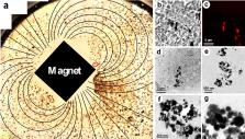
          Iron core nanoparticles with intact carbon shells were purified by magnetoseparation after hydrochloric acid treatment. The structure of the NPs (nanoparticles) was examined with a high resolution electron microscopy. The surface of the NPs was alkylcarboxylated and further aminated for covalent linking with Alexa Fluor 647 fluorochrome to produce modified fluorescent magnetic nanoparticles (MFMNPs). Live fluorescent imaging and correlative light-electron microscopy were used to study the NPs intracellular distribution and the effects of constant magnetic field on internalized NPs in the cell culture were analyzed. Cell viability was assayed by measuring a proliferative pool with Click-IT labeling.

          Results

          The microstructure and magnetic properties of superparamagnetic Fe@C core–shell NPs as well as their endocytosis by living tumor cells, and behavior inside the cells in constant magnetic field (150 mT) were studied. Correlative light-electron microscopy demonstrated that NPs retained their microstructure after internalization by the living cells. Application of constant magnetic field caused orientation of internalized NPs along power lines thus demonstrating their magnetocontrollability. Carbon onion-like shells make these NPs biocompatible and enable long-term observation with confocal microscope. It was found that iron core of NPs shows no toxic effect on the cell physiology, does not inhibit the cell proliferation and also does not induce apoptosis.

          Conclusions

          Non-toxic, biologically compatible superparamagnetic fluorescent MFMNPs can be further used for biological application such as delivery of biologically active compounds both inside the cell and inside the whole organism, magnetic separation, and magnetic resonance imaging (MRI) diagnostics.

          Related collections

          Most cited references26

          • Record: found
          • Abstract: found
          • Article: found
          Is Open Access

          Superparamagnetic iron oxide nanoparticles: magnetic nanoplatforms as drug carriers

          A targeted drug delivery system is the need of the hour. Guiding magnetic iron oxide nanoparticles with the help of an external magnetic field to its target is the principle behind the development of superparamagnetic iron oxide nanoparticles (SPIONs) as novel drug delivery vehicles. SPIONs are small synthetic γ-Fe2O3 (maghemite) or Fe3O4 (magnetite) particles with a core ranging between 10 nm and 100 nm in diameter. These magnetic particles are coated with certain biocompatible polymers, such as dextran or polyethylene glycol, which provide chemical handles for the conjugation of therapeutic agents and also improve their blood distribution profile. The current research on SPIONs is opening up wide horizons for their use as diagnostic agents in magnetic resonance imaging as well as for drug delivery vehicles. Delivery of anticancer drugs by coupling with functionalized SPIONs to their targeted site is one of the most pursued areas of research in the development of cancer treatment strategies. SPIONs have also demonstrated their efficiency as nonviral gene vectors that facilitate the introduction of plasmids into the nucleus at rates multifold those of routinely available standard technologies. SPION-induced hyperthermia has also been utilized for localized killing of cancerous cells. Despite their potential biomedical application, alteration in gene expression profiles, disturbance in iron homeostasis, oxidative stress, and altered cellular responses are some SPION-related toxicological aspects which require due consideration. This review provides a comprehensive understanding of SPIONs with regard to their method of preparation, their utility as drug delivery vehicles, and some concerns which need to be resolved before they can be moved from bench top to bedside.
            Bookmark
            • Record: found
            • Abstract: found
            • Article: not found

            Applications of nanoparticles in biology and medicine

            OV Salata (2004)
            Nanomaterials are at the leading edge of the rapidly developing field of nanotechnology. Their unique size-dependent properties make these materials superior and indispensable in many areas of human activity. This brief review tries to summarise the most recent developments in the field of applied nanomaterials, in particular their application in biology and medicine, and discusses their commercialisation prospects.
              Bookmark
              • Record: found
              • Abstract: found
              • Article: not found

              Strategies for the intracellular delivery of nanoparticles.

              The ability to target contrast agents and therapeutics inside cells is becoming important as we strive to decipher the complex network of events that occur within living cells and design therapies that can modulate these processes. Nanotechnology researchers have generated a growing list of nanoparticles designed for such applications. These particles can be assembled from a variety of materials into desirable geometries and configurations and possess useful properties and functionalities. Undoubtedly, the effective delivery of these nanomaterials into cells will be critical to their applications. In this tutorial review, we discuss the fundamental challenges of delivering nanoparticles into cells and to the targeted organelles, and summarize strategies that have been developed to-date.
                Bookmark

                Author and article information

                Contributors
                anastasiacit@gmail.com
                kireev@belozersky.msu.ru
                oxana.zhironkina@gmail.com
                olgastrelkova@gmail.com
                irina_alieva@belozersky.msu.ru
                valerydav@yandex.ru
                sankaran.murugesan@bakerhughes.com
                valery.khabashesku@bakerhughes.com
                alexander.majouga@gmail.com
                viatcheslav.agafonov@univ-tours.fr
                +33 247366070 , rustem.uzbekov@univ-tours.fr
                Journal
                J Nanobiotechnology
                J Nanobiotechnology
                Journal of Nanobiotechnology
                BioMed Central (London )
                1477-3155
                6 February 2019
                6 February 2019
                2019
                : 17
                : 27
                Affiliations
                [1 ]ISNI 0000 0001 2182 6141, GRID grid.12366.30, GREMAN, UMR CNRS 7347, , Université de Tours, ; 37200 Tours, France
                [2 ]ISNI 0000 0001 0010 3972, GRID grid.35043.31, National University of Science and Technology «MISiS», ; 119049 Moscow, Russian Federation
                [3 ]ISNI 0000 0001 2342 9668, GRID grid.14476.30, Faculty of Chemistry, , Lomonosov Moscow State University, ; 119991 Moscow, Russian Federation
                [4 ]ISNI 0000 0001 2342 9668, GRID grid.14476.30, Belozersky Institute of Physico-Chemical Biology, , Lomonosov Moscow State University, ; 119234 Moscow, Russian Federation
                [5 ]L. F. Vereshchagin Institute for High Pressure Physics of the RAS, 142190 Troitsk, Russian Federation
                [6 ]ISNI 0000 0004 0421 3468, GRID grid.467218.e, Center for Technology Innovation, , Baker Hughes a GE Company, ; Houston, TX 77040 USA
                [7 ]ISNI 0000 0004 0646 1385, GRID grid.39572.3a, D. Mendeleev University of Chemical Technology of Russia, ; Moscow, 125047 Russian Federation
                [8 ]ISNI 0000 0001 2182 6141, GRID grid.12366.30, Faculté de Médecine, , Université François Rabelais, ; 37032 Tours, France
                [9 ]ISNI 0000 0001 2342 9668, GRID grid.14476.30, Faculty of Bioengineering and Bioinformatics, , Lomonosov Moscow State University, ; 119192 Moscow, Russian Federation
                Author information
                http://orcid.org/0000-0002-9336-5484
                Article
                463
                10.1186/s12951-019-0463-5
                6364403
                30728022
                2582ca7d-393a-4d5f-9646-a6708f3a9dbf
                © The Author(s) 2019

                Open AccessThis article is distributed under the terms of the Creative Commons Attribution 4.0 International License ( http://creativecommons.org/licenses/by/4.0/), which permits unrestricted use, distribution, and reproduction in any medium, provided you give appropriate credit to the original author(s) and the source, provide a link to the Creative Commons license, and indicate if changes were made. The Creative Commons Public Domain Dedication waiver ( http://creativecommons.org/publicdomain/zero/1.0/) applies to the data made available in this article, unless otherwise stated.

                History
                : 5 November 2018
                : 29 January 2019
                Funding
                Funded by: FundRef http://dx.doi.org/10.13039/100002291, "Hyper diamond";
                Award ID: 667192
                Award Recipient :
                Categories
                Research
                Custom metadata
                © The Author(s) 2019

                Biotechnology
                superparamagnetic carbon-encapsulated iron nanoparticles,fluorescent nanoparticles,magnetocontrollability,human fibrosarcoma cell line,magnetic field,3-dimensional (3d) reconstruction,electron microscopy

                Comments

                Comment on this article