96
views
0
recommends
+1 Recommend
0 collections
    0
    shares
      • Record: found
      • Abstract: found
      • Article: found
      Is Open Access

      Defining Epidermal Basal Cell States during Skin Homeostasis and Wound Healing Using Single-Cell Transcriptomics

      research-article

      Read this article at

      Bookmark
          There is no author summary for this article yet. Authors can add summaries to their articles on ScienceOpen to make them more accessible to a non-specialist audience.

          SUMMARY

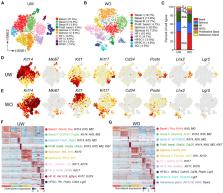
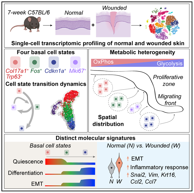
          Our knowledge of transcriptional heterogeneities in epithelial stem and progenitor cell compartments is limited. Epidermal basal cells sustain cutaneous tissue maintenance and drive wound healing. Previous studies have probed basal cell heterogeneity in stem and progenitor potential, but a comprehensive dissection of basal cell dynamics during differentiation is lacking. Using single-cell RNA sequencing coupled with RNAScope and fluorescence lifetime imaging, we identify three non-proliferative and one proliferative basal cell state in homeostatic skin that differ in metabolic preference and become spatially partitioned during wound re-epithelialization. Pseudotemporal trajectory and RNA velocity analyses predict a quasi-linear differentiation hierarchy where basal cells progress from Col17a1 Hi /Trp63 Hi state to early-response state, proliferate at the juncture of these two states, or become growth arrested before differentiating into spinous cells. Wound healing induces plasticity manifested by dynamic basal-spinous interconversions at multiple basal transcriptional states. Our study provides a systematic view of epidermal cellular dynamics, supporting a revised “hierarchical-lineage” model of homeostasis.

          Graphical Abstract

          In Brief

          Haensel et al. performed a comprehensive dissection of the cellular makeup of skin during homeostasis and wound healing and the molecular heterogeneity and cellular dynamics within its stem-cell-containing epidermal basal layer. Their work provides insights and stimulates further investigation into the mechanism of skin maintenance and repair.

          Related collections

          Most cited references61

          • Record: found
          • Abstract: found
          • Article: not found

          p63 is essential for regenerative proliferation in limb, craniofacial and epithelial development.

          The p63 gene, a homologue of the tumour-suppressor p53, is highly expressed in the basal or progenitor layers of many epithelial tissues. Here we report that mice homozygous for a disrupted p63 gene have major defects in their limb, craniofacial and epithelial development. p63 is expressed in the ectodermal surfaces of the limb buds, branchial arches and epidermal appendages, which are all sites of reciprocal signalling that direct morphogenetic patterning of the underlying mesoderm. The limb truncations are due to a failure to maintain the apical ectodermal ridge, a stratified epithelium, essential for limb development. The embryonic epidermis of p63-/- mice undergoes an unusual process of non-regenerative differentiation, culminating in a striking absence of all squamous epithelia and their derivatives, including mammary, lacrymal and salivary glands. Taken together, our results indicate that p63 is critical for maintaining the progenitor-cell populations that are necessary to sustain epithelial development and morphogenesis.
            Bookmark
            • Record: found
            • Abstract: found
            • Article: not found

            Molecular regulation of stem cell quiescence.

            Subsets of mammalian adult stem cells reside in the quiescent state for prolonged periods of time. This state, which is reversible, has long been viewed as dormant and with minimal basal activity. Recent advances in adult stem cell isolation have provided insights into the epigenetic, transcriptional and post-transcriptional control of quiescence and suggest that quiescence is an actively maintained state in which signalling pathways are involved in maintaining a poised state that allows rapid activation. Deciphering the molecular mechanisms regulating adult stem cell quiescence will increase our understanding of tissue regeneration mechanisms and how they are dysregulated in pathological conditions and in ageing.
              Bookmark
              • Record: found
              • Abstract: found
              • Article: not found

              p63 is a p53 homologue required for limb and epidermal morphogenesis.

              The p53 tumour suppressor is a transcription factor that regulates the progression of the cell through its cycle and cell death (apoptosis) in response to environmental stimuli such as DNA damage and hypoxia. Even though p53 modulates these critical cellular processes, mice that lack p53 are developmentally normal, suggesting that p53-related proteins might compensate for the functions of p53 during embryogenesis. Two p53 homologues, p63 and p73, are known and here we describe the function of p63 in vivo. Mice lacking p63 are born alive but have striking developmental defects. Their limbs are absent or truncated, defects that are caused by a failure of the apical ectodermal ridge to differentiate. The skin of p63-deficient mice does not progress past an early developmental stage: it lacks stratification and does not express differentiation markers. Structures dependent upon epidermal-mesenchymal interactions during embryonic development, such as hair follicles, teeth and mammary glands, are absent in p63-deficient mice. Thus, in contrast to p53, p63 is essential for several aspects of ectodermal differentiation during embryogenesis.
                Bookmark

                Author and article information

                Journal
                101573691
                39703
                Cell Rep
                Cell Rep
                Cell reports
                2211-1247
                27 March 2020
                17 March 2020
                13 May 2020
                : 30
                : 11
                : 3932-3947.e6
                Affiliations
                [1 ]Department of Biological Chemistry, School of Medicine, University of California, Irvine, CA 92697, USA
                [2 ]The NSF-Simons Center for Multiscale Cell Fate Research, University of California, Irvine, CA 92627, USA
                [3 ]Department of Mathematics, University of California, Irvine, CA 92697, USA
                [4 ]Laboratory for Fluorescence Dynamics, Department of Biomedical Engineering, University of California, Irvine, CA 92697, USA
                [5 ]Center for Complex Biological Systems, University of California, Irvine, CA 92697, USA
                [6 ]Department of Developmental and Cell Biology, University of California, Irvine, CA 92697, USA
                [7 ]These authors contributed equally
                [8 ]Lead Contact
                Author notes
                [* ]Correspondence: qnie@ 123456uci.edu (Q.N.), xdai@ 123456uci.edu (X.D.)

                AUTHOR CONTRIBUTIONS

                X.D. and D.H. conceived the study and designed the experiments; X.D. directed the project; R.C. performed FLIM analysis under E.G.’s supervision; Q. Nguyen assisted with sample processing and sequencing under K.K.’s supervision; and D.H., P.S., M.D., and R.V. performed the remaining wet-lab experiments under X.D.’s supervision. D.H., S.J., Z.C., Q. Nguyen, Y.G., and A.L.M. performed or assisted with the computational analysis under X.D.’s and Q. Nie’s supervision; D.H., S.J., and X.D. wrote the manuscript with input from all authors.

                Article
                NIHMS1578466
                10.1016/j.celrep.2020.02.091
                7218802
                32187560
                292def9b-3346-4030-8331-39327a00db63

                This is an open access article under the CC BY-NC-ND license ( http://creativecommons.org/licenses/by-nc-nd/4.0/).

                History
                Categories
                Article

                Cell biology
                Cell biology

                Comments

                Comment on this article