2
views
0
recommends
+1 Recommend
0 collections
    0
    shares
      • Record: found
      • Abstract: found
      • Article: found
      Is Open Access

      Oncogenic Vav1-Myo1f induces therapeutically targetable macrophage-rich tumor microenvironment in peripheral T cell lymphoma

      research-article

      Read this article at

      Bookmark
          There is no author summary for this article yet. Authors can add summaries to their articles on ScienceOpen to make them more accessible to a non-specialist audience.

          SUMMARY

          Peripheral T cell lymphoma not otherwise specified (PTCL-NOS) comprises heterogeneous lymphoid malignancies characterized by pleomorphic lymphocytes and variable inflammatory cell-rich tumor microenvironment. Genetic drivers in PTCL-NOS include genomic alterations affecting the VAV1 oncogene; however, their specific role and mechanisms in PTCL-NOS remain incompletely understood. Here we show that expression of Vav1-Myo1f, a recurrent PTCL-associated VAV1 fusion, induces oncogenic transformation of CD4 + T cells. Notably, mouse Vav1-Myo1f lymphomas show T helper type 2 features analogous to high-risk GATA3 + human PTCL. Single-cell transcriptome analysis reveals that Vav1-Myo1f alters T cell differentiation and leads to accumulation of tumor-associated macrophages (TAMs) in the tumor microenvironment, a feature linked with aggressiveness in human PTCL. Importantly, therapeutic targeting of TAMs induces strong anti-lymphoma effects, highlighting the lymphoma cells’ dependency on the microenvironment. These results demonstrate an oncogenic role for Vav1-Myo1f in the pathogenesis of PTCL, involving deregulation in T cell polarization, and identify the lymphoma-associated macrophage-tumor microenvironment as a therapeutic target in PTCL.
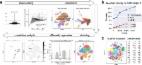
          Graphical Abstract

          In brief

          Cortes et al. show that expression of Vav1-Myo1f, a recurrent peripheral T cell lymphoma (PTCL)-associated VAV1 fusion, induces CD4 + T cell lymphoma with features analogous to high-risk GATA3 + human PTCL. Expression of Vav1-Myo1f induces recruitment of tumor-associated macrophages to the tumor microenvironment that can be targeted for therapeutic intervention in PTCL.

          Related collections

          Most cited references61

          • Record: found
          • Abstract: found
          • Article: not found

          Gene set enrichment analysis: A knowledge-based approach for interpreting genome-wide expression profiles

          Although genomewide RNA expression analysis has become a routine tool in biomedical research, extracting biological insight from such information remains a major challenge. Here, we describe a powerful analytical method called Gene Set Enrichment Analysis (GSEA) for interpreting gene expression data. The method derives its power by focusing on gene sets, that is, groups of genes that share common biological function, chromosomal location, or regulation. We demonstrate how GSEA yields insights into several cancer-related data sets, including leukemia and lung cancer. Notably, where single-gene analysis finds little similarity between two independent studies of patient survival in lung cancer, GSEA reveals many biological pathways in common. The GSEA method is embodied in a freely available software package, together with an initial database of 1,325 biologically defined gene sets.
            Bookmark
            • Record: found
            • Abstract: found
            • Article: not found

            Integrating single-cell transcriptomic data across different conditions, technologies, and species

            Computational single-cell RNA-seq (scRNA-seq) methods have been successfully applied to experiments representing a single condition, technology, or species to discover and define cellular phenotypes. However, identifying subpopulations of cells that are present across multiple data sets remains challenging. Here, we introduce an analytical strategy for integrating scRNA-seq data sets based on common sources of variation, enabling the identification of shared populations across data sets and downstream comparative analysis. We apply this approach, implemented in our R toolkit Seurat (http://satijalab.org/seurat/), to align scRNA-seq data sets of peripheral blood mononuclear cells under resting and stimulated conditions, hematopoietic progenitors sequenced using two profiling technologies, and pancreatic cell 'atlases' generated from human and mouse islets. In each case, we learn distinct or transitional cell states jointly across data sets, while boosting statistical power through integrated analysis. Our approach facilitates general comparisons of scRNA-seq data sets, potentially deepening our understanding of how distinct cell states respond to perturbation, disease, and evolution.
              Bookmark
              • Record: found
              • Abstract: found
              • Article: found
              Is Open Access

              SCANPY : large-scale single-cell gene expression data analysis

              Scanpy is a scalable toolkit for analyzing single-cell gene expression data. It includes methods for preprocessing, visualization, clustering, pseudotime and trajectory inference, differential expression testing, and simulation of gene regulatory networks. Its Python-based implementation efficiently deals with data sets of more than one million cells (https://github.com/theislab/Scanpy). Along with Scanpy, we present AnnData, a generic class for handling annotated data matrices (https://github.com/theislab/anndata).
                Bookmark

                Author and article information

                Journal
                101573691
                39703
                Cell Rep
                Cell Rep
                Cell reports
                2211-1247
                22 April 2022
                19 April 2022
                02 May 2022
                : 39
                : 3
                : 110695
                Affiliations
                [1 ]Institute for Cancer Genetics, Columbia University, New York, NY 10032, USA
                [2 ]Department of Systems Biology, Columbia University, New York, NY 10032, USA
                [3 ]Department of Pathology and Cell Biology, Columbia University, New York, NY 10032, USA
                [4 ]Department of Biomedical Informatics, Columbia University, New York, NY 10032, USA
                [5 ]Department of Hematology, Hospital Clínic de Barcelona, Barcelona 08036, Spain
                [6 ]Experimental Oncology Laboratory, IRCCS Istituto Ortopedico Rizzoli, Bologna 40136, Italy
                [7 ]Division of Hematopathology, European Institute of Oncology IRCCS, Milan 20141, Italy
                [8 ]Hematopathology Unit, Department of Pathology, Hospital Clínic-IDIBAPS, Barcelona 08036, Spain
                [9 ]Department of Pediatrics, Columbia University, New York, NY 10032, USA
                [10 ]These authors contributed equally
                [11 ]Lead contact
                Author notes

                AUTHOR CONTRIBUTIONS

                Conception and design, J.R.C. and T.P.; Development of methodology, J.R.C., R.A., I.F., J.A.P.-G., and S.A.Q.; Acquisition of data (provided animals, performed experiments, analyzed experiments, provided histopathological analysis and acquired human samples), J.R.C., R.A., W.-H.W.L., A.P.L., B.B.S., J.A.B., A.J.C., A.R.-G., M.A.L., S.P., E.C., and G.B.; Analysis and interpretation of data (e.g., statistical analysis, biostatistics, computational analysis), J.R.C., R.A., I.F., J.A.P.-G., and S.A.Q.; Writing, review, and/or revision of the manuscript, J.R.C., A.A.F., and T.P.; Administrative, technical, or material support (i.e., reporting or organizing data, constructing databases), A.J.C., A.M., J.E., and S.Z.; Study supervision, J.R.C., R.R., A.A.F., and T.P.

                [* ]Correspondence: tp2151@ 123456columbia.edu
                Article
                NIHMS1799981
                10.1016/j.celrep.2022.110695
                9059228
                35443168
                3bfd5646-31d3-46ed-bac2-b0d27efbda5a

                This is an open access article under the CC BY-NC-ND license ( http://creativecommons.org/licenses/by-nc-nd/4.0/).

                History
                Categories
                Article

                Cell biology
                Cell biology

                Comments

                Comment on this article