20
views
0
recommends
+1 Recommend
0 collections
    0
    shares
      • Record: found
      • Abstract: found
      • Article: found
      Is Open Access

      Expression of human eukaryotic initiation factor 3f oscillates with cell cycle in A549 cells and is essential for cell viability

      research-article
      1 , 2 ,
      Cell Division
      BioMed Central

      Read this article at

      Bookmark
          There is no author summary for this article yet. Authors can add summaries to their articles on ScienceOpen to make them more accessible to a non-specialist audience.

          Abstract

          Background

          Transcriptional and postranslational regulation of the cell cycle has been widely studied. However, there is scarce knowledge concerning translational control of this process. Several mammalian eukaryotic initiation factors (eIFs) seem to be implicated in controlling cell proliferation. In this work, we investigated if the human eIF3f expression and function is cell cycle related.

          Results

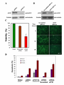
          The human eIF3f expression has been found to be upregulated in growth-stimulated A549 cells and downregulated in G0. Western blot analysis and eIF3f promotor-luciferase fusions revealed that eIF3f expression peaks twice in the cell cycle: in the S and the M phases. Deregulation of eIF3f expression negatively affects cell viability and induces apoptosis.

          Conclusions

          The expression pattern of human eIF3f during the cell cycle confirms that this gene is cell division related. The fact that eIF3f expression peaks in two cell cycle phases raises the possibility that this gene may exert a differential function in the S and M phases. Our results strongly suggest that eIF3f is essential for cell proliferation.

          Related collections

          Most cited references29

          • Record: found
          • Abstract: found
          • Article: not found

          Evolutionary fate of retroposed gene copies in the human genome.

          Given that retroposed copies of genes are presumed to lack the regulatory elements required for their expression, retroposition has long been considered a mechanism without functional relevance. However, through an in silico assay for transcriptional activity, we identify here >1,000 transcribed retrocopies in the human genome, of which at least approximately 120 have evolved into bona fide genes. Among these, approximately 50 retrogenes have evolved functions in testes, more than half of which were recruited as functional autosomal counterparts of X-linked genes during spermatogenesis. Generally, retrogenes emerge "out of the testis," because they are often initially transcribed in testis and later evolve stronger and sometimes more diverse spatial expression patterns. We find a significant excess of transcribed retrocopies close to other genes or within introns, suggesting that retrocopies can exploit the regulatory elements and/or open chromatin of neighboring genes to become transcribed. In direct support of this hypothesis, we identify 36 retrocopy-host gene fusions, including primate-specific chimeric genes. Strikingly, 27 intergenic retrogenes have acquired untranslated exons de novo during evolution to achieve high expression levels. Notably, our screen for highly transcribed retrocopies also uncovered a retrogene linked to a human recessive disorder, gelatinous drop-like corneal dystrophy, a form of blindness. These functional implications for retroposition notwithstanding, we find that the insertion of retrocopies into genes is generally deleterious, because it may interfere with the transcription of host genes. Our results demonstrate that natural selection has been fundamental in shaping the retrocopy repertoire of the human genome.
            Bookmark
            • Record: found
            • Abstract: found
            • Article: not found

            Therapeutic suppression of translation initiation factor eIF4E expression reduces tumor growth without toxicity.

            Expression of eukaryotic translation initiation factor 4E (eIF4E) is commonly elevated in human and experimental cancers, promoting angiogenesis and tumor growth. Elevated eIF4E levels selectively increase translation of growth factors important in malignancy (e.g., VEGF, cyclin D1) and is thereby an attractive anticancer therapeutic target. Yet to date, no eIF4E-specific therapy has been developed. Herein we report development of eIF4E-specific antisense oligonucleotides (ASOs) designed to have the necessary tissue stability and nuclease resistance required for systemic anticancer therapy. In mammalian cultured cells, these ASOs specifically targeted the eIF4E mRNA for destruction, repressing expression of eIF4E-regulated proteins (e.g., VEGF, cyclin D1, survivin, c-myc, Bcl-2), inducing apoptosis, and preventing endothelial cells from forming vessel-like structures. Most importantly, intravenous ASO administration selectively and significantly reduced eIF4E expression in human tumor xenografts, significantly suppressing tumor growth. Because these ASOs also target murine eIF4E, we assessed the impact of eIF4E reduction in normal tissues. Despite reducing eIF4E levels by 80% in mouse liver, eIF4E-specific ASO administration did not affect body weight, organ weight, or liver transaminase levels, thereby providing the first in vivo evidence that cancers may be more susceptible to eIF4E inhibition than normal tissues. These data have prompted eIF4E-specific ASO clinical trials for the treatment of human cancers.
              Bookmark
              • Record: found
              • Abstract: found
              • Article: not found

              Mammalian retroelements.

              The eukaryotic genome has undergone a series of epidemics of amplification of mobile elements that have resulted in most eukaryotic genomes containing much more of this 'junk' DNA than actual coding DNA. The majority of these elements utilize an RNA intermediate and are termed retroelements. Most of these retroelements appear to amplify in evolutionary waves that insert in the genome and then gradually diverge. In humans, almost half of the genome is recognizably derived from retroelements, with the two elements that are currently actively amplifying, L1 and Alu, making up about 25% of the genome and contributing extensively to disease. The mechanisms of this amplification process are beginning to be understood, although there are still more questions than answers. Insertion of new retroelements may directly damage the genome, and the presence of multiple copies of these elements throughout the genome has longer-term influences on recombination events in the genome and more subtle influences on gene expression.
                Bookmark

                Author and article information

                Journal
                Cell Div
                Cell Division
                BioMed Central
                1747-1028
                2010
                13 May 2010
                : 5
                : 10
                Affiliations
                [1 ]División de Estudios de Posgrado, Facultad de Ciencias Médicas y Biológicas Dr. Ignacio Chávez, Universidad Michoacana de San Nicolás de Hidalgo, Morelia, Michoacán, México
                [2 ]Departmento de Biología Molecular, Instituto de Investigaciones Químico-Biológicas, Universidad Michoacana de San Nicolás de Hidalgo, Morelia, Michoacán, México
                Article
                1747-1028-5-10
                10.1186/1747-1028-5-10
                2877012
                20462454
                45c50628-abc2-4836-ae08-0ab82dd5363c
                Copyright ©2010 Higareda-Mendoza and Pardo-Galván; licensee BioMed Central Ltd.

                This is an Open Access article distributed under the terms of the Creative Commons Attribution License ( http://creativecommons.org/licenses/by/2.0), which permits unrestricted use, distribution, and reproduction in any medium, provided the original work is properly cited.

                History
                : 22 October 2009
                : 13 May 2010
                Categories
                Research

                Cell biology
                Cell biology

                Comments

                Comment on this article