14
views
0
recommends
+1 Recommend
0 collections
    0
    shares
      • Record: found
      • Abstract: found
      • Article: not found

      Regulation of Calreticulin Gene Expression by Calcium

      research-article

      Read this article at

      ScienceOpenPublisherPMC
      Bookmark
          There is no author summary for this article yet. Authors can add summaries to their articles on ScienceOpen to make them more accessible to a non-specialist audience.

          Abstract

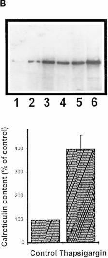
          We have isolated and characterized a 12-kb mouse genomic DNA fragment containing the entire calreticulin gene and 2.14 kb of the promoter region. The mouse calreticulin gene consists of nine exons and eight introns, and it spans 4.2 kb of genomic DNA. A 1.8-kb fragment of the calreticulin promoter was subcloned into a reporter gene plasmid containing chloramphenicol acetyltransferase. This construct was then used in transient and stable transfection of NIH/ 3T3 cells. Treatment of transfected cells either with the Ca 2+ ionophore A23187, or with the ER Ca 2+-ATPase inhibitor thapsigargin, resulted in a five- to sevenfold increase of the expression of chloramphenicol acetyltransferase protein. Transactivation of the calreticulin promoter was also increased by fourfold in NIH/3T3 cells treated with bradykinin, a hormone that induces Ca 2+ release from the intracellular Ca 2+ stores. Analysis of the promoter deletion constructs revealed that A23187- and thapsigargin-responsive regions are confined to two regions (−115 to −260 and −685 to −1,763) in the calreticulin promoter that contain the CCAAT nucleotide sequences. Northern blot analysis of cells treated with A23187, or with thapsigargin, revealed a fivefold increase in calreticulin mRNA levels. Thapsigargin also induced a fourfold increase in calreticulun protein levels. Importantly, we show by nuclear run-on transcription analysis that calreticulin gene transcription is increased in NIH/3T3 cells treated with A23187 and thapsigargin in vivo. This increase in gene expression required over 4 h of continuous incubation with the drugs and was also sensitive to treatment with cycloheximide, suggesting that it is dependent on protein synthesis. Changes in the concentration of extracellular and cytoplasmic Ca 2+ did not affect the increased expression of the calreticulin gene. These studies suggest that stress response to the depletion of intracellular Ca 2+ stores induces expression of the calreticulin gene in vitro and in vivo.

          Related collections

          Most cited references70

          • Record: found
          • Abstract: found
          • Article: not found

          Protein folding in the cell.

          In the cell, as in vitro, the final conformation of a protein is determined by its amino-acid sequence. But whereas some isolated proteins can be denatured and refolded in vitro in the absence of other macromolecular cellular components, folding and assembly of polypeptides in vivo involves other proteins, many of which belong to families that have been highly conserved during evolution.
            Bookmark
            • Record: found
            • Abstract: found
            • Article: not found

            Transcriptional induction of genes encoding endoplasmic reticulum resident proteins requires a transmembrane protein kinase.

            The transcription of genes encoding soluble proteins that reside in the endoplasmic reticulum (ER) is induced when unfolded proteins accumulate in the ER. Thus, an intracellular signal transduction pathway must exist that mediates communication between the ER lumen and the nucleus. We have identified a gene in S. cerevisiae, IRE1, that is required for this pathway: ire1- mutants cannot activate transcription of KAR2 and PDI1, which encode the ER resident proteins BiP and protein disulfide isomerase. Moreover, IRE1 is essential for cell viability under stress conditions that cause unfolded proteins to accumulate in the ER. IRE1 encodes a transmembrane serine/threonine kinase that we propose transmits the unfolded protein signal across the ER or inner nuclear membrane. IRE1 is also required for inositol prototrophy, suggesting that the induction of ER resident proteins is coupled to the biogenesis of new ER membrane.
              Bookmark
              • Record: found
              • Abstract: found
              • Article: not found

              A transmembrane protein with a cdc2+/CDC28-related kinase activity is required for signaling from the ER to the nucleus.

              In eukaryotic cells, the accumulation of unfolded proteins in the endoplasmic reticulum (ER) triggers a signaling pathway from the ER to the nucleus. Several yeast mutants defective in this pathway map to the ERN1 gene, which protects cells from lethal consequences of stress by signaling for increased expression of BiP and other ER proteins. ERN1 encodes a 1115 amino acid transmembrane protein (Ern1p) whose glycosylated N-terminal portion is located inside microsomes and whose cytoplasmic C-terminal portion carries an essential protein kinase activity. We postulate that Ern1p is the proximal sensor of events in the ER and that binding of ligand causes transduction of information across the ER membrane, leading to activation of a specific set of transcription factors.
                Bookmark

                Author and article information

                Journal
                J Cell Biol
                The Journal of Cell Biology
                The Rockefeller University Press
                0021-9525
                1540-8140
                11 August 1997
                : 138
                : 3
                : 547-557
                Affiliations
                Medical Research Council Group in Molecular Biology of Membranes, Department of Biochemistry, University of Alberta, Edmonton, Alberta, Canada T6G 2S2
                Article
                10.1083/jcb.138.3.547
                2141645
                9245785
                4e04c4ab-419b-4a15-ae17-d8ce97c0b1fd
                Copyright @ 1997
                History
                : 20 November 1996
                : 11 March 1997
                Categories
                Article

                Cell biology
                Cell biology

                Comments

                Comment on this article