9
views
0
recommends
+1 Recommend
0 collections
    0
    shares
      • Record: found
      • Abstract: found
      • Article: found
      Is Open Access

      Iron-induced cytotoxicity mediated by endolysosomal TRPML1 channels is reverted by TFEB

      research-article

      Read this article at

      Bookmark
          There is no author summary for this article yet. Authors can add summaries to their articles on ScienceOpen to make them more accessible to a non-specialist audience.

          Abstract

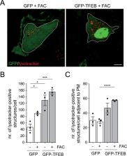
          Increased brain iron content has been consistently reported in sporadic Parkinson’s disease (PD) patients, and an increase in cytosolic free iron is known to cause oxidative stress and cell death. However, whether iron also accumulates in susceptible brain areas in humans or in mouse models of familial PD remains unknown. In addition, whilst the lysosome functions as a critical intracellular iron storage organelle, little is known about the mechanisms underlying lysosomal iron release and how this process is influenced by lysosome biogenesis and/or lysosomal exocytosis. Here, we report an increase in brain iron content also in PD patients due to the common G2019S-LRRK2 mutation as compared to healthy age-matched controls, whilst differences in iron content are not observed in G2019S-LRRK2 knockin as compared to control mice. Chemically triggering iron overload in cultured cells causes cytotoxicity via the endolysosomal release of iron which is mediated by TRPML1. TFEB expression reverts the iron overload-associated cytotoxicity by causing lysosomal exocytosis, which is dependent on a TRPML1-mediated increase in cytosolic calcium levels. Therefore, approaches aimed at increasing TFEB levels, or pharmacological TRPML1 activation in conjunction with iron chelation may prove beneficial against cell death associated with iron overload conditions such as those associated with PD.

          Related collections

          Most cited references66

          • Record: found
          • Abstract: found
          • Article: not found

          A gene network regulating lysosomal biogenesis and function.

          Lysosomes are organelles central to degradation and recycling processes in animal cells. Whether lysosomal activity is coordinated to respond to cellular needs remains unclear. We found that most lysosomal genes exhibit coordinated transcriptional behavior and are regulated by the transcription factor EB (TFEB). Under aberrant lysosomal storage conditions, TFEB translocated from the cytoplasm to the nucleus, resulting in the activation of its target genes. TFEB overexpression in cultured cells induced lysosomal biogenesis and increased the degradation of complex molecules, such as glycosaminoglycans and the pathogenic protein that causes Huntington's disease. Thus, a genetic program controls lysosomal biogenesis and function, providing a potential therapeutic target to enhance cellular clearing in lysosomal storage disorders and neurodegenerative diseases.
            Bookmark
            • Record: found
            • Abstract: found
            • Article: not found

            The role of iron in brain ageing and neurodegenerative disorders.

            In the CNS, iron in several proteins is involved in many important processes such as oxygen transportation, oxidative phosphorylation, myelin production, and the synthesis and metabolism of neurotransmitters. Abnormal iron homoeostasis can induce cellular damage through hydroxyl radical production, which can cause the oxidation and modification of lipids, proteins, carbohydrates, and DNA. During ageing, different iron complexes accumulate in brain regions associated with motor and cognitive impairment. In various neurodegenerative diseases, such as Alzheimer's disease and Parkinson's disease, changes in iron homoeostasis result in altered cellular iron distribution and accumulation. MRI can often identify these changes, thus providing a potential diagnostic biomarker of neurodegenerative diseases. An important avenue to reduce iron accumulation is the use of iron chelators that are able to cross the blood-brain barrier, penetrate cells, and reduce excessive iron accumulation, thereby affording neuroprotection.
              Bookmark
              • Record: found
              • Abstract: found
              • Article: not found

              Lysosomal calcium signaling regulates autophagy via calcineurin and TFEB

              The view of the lysosome as the terminal end of cellular catabolic pathways has been challenged by recent studies showing a central role of this organelle in the control of cell function. Here we show that a lysosomal Ca2+ signaling mechanism controls the activities of the phosphatase calcineurin and of its substrate TFEB, a master transcriptional regulator of lysosomal biogenesis and autophagy. Lysosomal Ca2+ release via mucolipin 1 (MCOLN1) activates calcineurin, which binds and de-phosphorylates TFEB, thus promoting its nuclear translocation. Genetic and pharmacological inhibition of calcineurin suppressed TFEB activity during starvation and physical exercise, while calcineurin overexpression and constitutive activation had the opposite effect. Induction of autophagy and lysosomal biogenesis via TFEB required MCOLN1-mediated calcineurin activation, linking lysosomal calcium signaling to both calcineurin regulation and autophagy induction. Thus, the lysosome reveals itself as a hub for the signaling pathways that regulate cellular homeostasis.
                Bookmark

                Author and article information

                Contributors
                sabine.hilfiker@rutgers.edu
                Journal
                Cell Death Dis
                Cell Death Dis
                Cell Death & Disease
                Nature Publishing Group UK (London )
                2041-4889
                16 December 2022
                16 December 2022
                December 2022
                : 13
                : 12
                : 1047
                Affiliations
                [1 ]GRID grid.4711.3, ISNI 0000 0001 2183 4846, Institute of Parasitology and Biomedicine “López-Neyra”, , Consejo Superior de Investigaciones Científicas (CSIC), ; 18016 Granada, Spain
                [2 ]Department of Legal Medicine and Toxicology, School of Medicine, University of 18016 Granada, Granada, Spain
                [3 ]GRID grid.430387.b, ISNI 0000 0004 1936 8796, Department of Anesthesiology and Department of Physiology, Pharmacology and Neuroscience, , Rutgers New Jersey Medical School, ; Newark, NJ 07103 USA
                [4 ]GRID grid.214458.e, ISNI 0000000086837370, Life Sciences Institute and Department of Cell and Developmental Biology, , University of Michigan, ; Ann Arbor, MI 48109 USA
                [5 ]GRID grid.5252.0, ISNI 0000 0004 1936 973X, Department of Pharmacy – Center for Drug Research, , Ludwig-Maximilians-University, ; 81377 Munich, Germany
                [6 ]GRID grid.5252.0, ISNI 0000 0004 1936 973X, Walther Straub Institute of Pharmacology and Toxicology Faculty of Medicine, , Ludwig-Maximilians-Universitaet, ; 80336 Munich, Germany
                [7 ]GRID grid.4991.5, ISNI 0000 0004 1936 8948, Department of Pharmacology, , University of Oxford, ; Oxford, OX1 3QT UK
                Author information
                http://orcid.org/0000-0001-9987-1495
                http://orcid.org/0000-0002-0177-5559
                Article
                5504
                10.1038/s41419-022-05504-2
                9755144
                36522443
                5fbfe993-d8a2-4f53-ba98-2497f351cc3c
                © The Author(s) 2022

                Open Access This article is licensed under a Creative Commons Attribution 4.0 International License, which permits use, sharing, adaptation, distribution and reproduction in any medium or format, as long as you give appropriate credit to the original author(s) and the source, provide a link to the Creative Commons license, and indicate if changes were made. The images or other third party material in this article are included in the article’s Creative Commons license, unless indicated otherwise in a credit line to the material. If material is not included in the article’s Creative Commons license and your intended use is not permitted by statutory regulation or exceeds the permitted use, you will need to obtain permission directly from the copyright holder. To view a copy of this license, visit http://creativecommons.org/licenses/by/4.0/.

                History
                : 19 October 2022
                : 30 November 2022
                : 7 December 2022
                Funding
                Funded by: CSIC (intramural funding) Rutgers University (intramural funding)
                Categories
                Article
                Custom metadata
                © The Author(s) 2022

                Cell biology
                mechanisms of disease,cell biology
                Cell biology
                mechanisms of disease, cell biology

                Comments

                Comment on this article