36
views
0
recommends
+1 Recommend
0 collections
    0
    shares
      • Record: found
      • Abstract: found
      • Article: found
      Is Open Access

      Non-invasive Imaging and Modeling of Liver Regeneration After Partial Hepatectomy

      research-article

      Read this article at

      Bookmark
          There is no author summary for this article yet. Authors can add summaries to their articles on ScienceOpen to make them more accessible to a non-specialist audience.

          Abstract

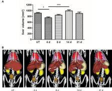
          The liver has a unique regenerative capability upon injury or partial resection. The regeneration process comprises a complex interplay between parenchymal and non-parenchymal cells and is tightly regulated at different scales. Thus, we investigated liver regeneration using multi-scale methods by combining non-invasive imaging with immunohistochemical analyses. In this context, non-invasive imaging can provide quantitative data of processes involved in liver regeneration at organ and body scale. We quantitatively measured liver volume recovery after 70% partial hepatectomy (PHx) by micro computed tomography (μCT) and investigated changes in the density of CD68 + macrophages by fluorescence-mediated tomography (FMT) combined with μCT using a newly developed near-infrared fluorescent probe. In addition, angiogenesis and tissue-resident macrophages were analyzed by immunohistochemistry. Based on the results, a model describing liver regeneration and the interactions between different cell types was established. In vivo analysis of liver volume regeneration over 21 days after PHx by μCT imaging demonstrated that the liver volume rapidly increased after PHx reaching a maximum at day 14 and normalizing until day 21. An increase in CD68 + macrophage density in the liver was detected from day 4 to day 8 by combined FMT-μCT imaging, followed by a decline towards control levels between day 14 and day 21. Immunohistochemistry revealed the highest angiogenic activity at day 4 after PHx that continuously declined thereafter, whereas the density of tissue-resident CD169 + macrophages was not altered. The simulated time courses for volume recovery, angiogenesis and macrophage density reflect the experimental data describing liver regeneration after PHx at organ and tissue scale. In this context, our study highlights the importance of non-invasive imaging for acquiring quantitative organ scale data that enable modeling of liver regeneration.

          Related collections

          Most cited references24

          • Record: found
          • Abstract: found
          • Article: not found

          Liver regeneration: from myth to mechanism.

          The unusual regenerative properties of the liver are a logical adaptation by organisms, as the liver is the main detoxifying organ of the body and is likely to be injured by ingested toxins. The numerous cytokine- and growth-factor-mediated pathways that are involved in regulating liver regeneration are being successfully dissected using molecular and genetic approaches. So what is known about this process at present and which questions remain?
            Bookmark
            • Record: found
            • Abstract: found
            • Article: not found

            Liver regeneration after partial hepatectomy: critical analysis of mechanistic dilemmas.

            Liver regeneration after partial hepatectomy is one of the most studied models of cell, organ, and tissue regeneration. The complexity of the signaling pathways initiating and terminating this process have provided paradigms for regenerative medicine. Many aspects of the signaling mechanisms involved in hepatic regeneration are under active investigation. The purpose of this review is to focus on the areas still not well understood. The review also aims to provide insights into the ways by which current concepts of liver regeneration can provide understanding regarding malfunction of the regenerative process in liver diseases, such as acute liver failure.
              Bookmark
              • Record: found
              • Abstract: found
              • Article: not found

              Prediction and validation of cell alignment along microvessels as order principle to restore tissue architecture in liver regeneration.

              Only little is known about how cells coordinately behave to establish functional tissue structure and restore microarchitecture during regeneration. Research in this field is hampered by a lack of techniques that allow quantification of tissue architecture and its development. To bridge this gap, we have established a procedure based on confocal laser scans, image processing, and three-dimensional tissue reconstruction, as well as quantitative mathematical modeling. As a proof of principle, we reconstructed and modeled liver regeneration in mice after damage by CCl(4), a prototypical inducer of pericentral liver damage. We have chosen the regenerating liver as an example because of the tight link between liver architecture and function: the complex microarchitecture formed by hepatocytes and microvessels, i.e. sinusoids, ensures optimal exchange of metabolites between blood and hepatocytes. Our model captures all hepatocytes and sinusoids of a liver lobule during a 16 days regeneration process. The model unambiguously predicted a so-far unrecognized mechanism as essential for liver regeneration, whereby daughter hepatocytes align along the orientation of the closest sinusoid, a process which we named "hepatocyte-sinusoid alignment" (HSA). The simulated tissue architecture was only in agreement with the experimentally obtained data when HSA was included into the model and, moreover, no other likely mechanism could replace it. In order to experimentally validate the model of prediction of HSA, we analyzed the three-dimensional orientation of daughter hepatocytes in relation to the sinusoids. The results of this analysis clearly confirmed the model prediction. We believe our procedure is widely applicable in the systems biology of tissues.
                Bookmark

                Author and article information

                Contributors
                Journal
                Front Physiol
                Front Physiol
                Front. Physiol.
                Frontiers in Physiology
                Frontiers Media S.A.
                1664-042X
                17 July 2019
                2019
                : 10
                : 904
                Affiliations
                [1] 1Institute for Experimental Molecular Imaging, Medical Faculty, RWTH Aachen University , Aachen, Germany
                [2] 2Department of Gastroenterology, Hepatology and Infectious Diseases, Medical Faculty, Heinrich-Heine-University Düsseldorf , Düsseldorf, Germany
                [3] 3Fraunhofer Institute for Molecular Biology and Applied Ecology IME , Aachen, Germany
                [4] 4Institute for Molecular Biotechnology, RWTH Aachen University , Aachen, Germany
                Author notes

                Edited by: Andreas Teufel, University of Heidelberg, Germany

                Reviewed by: Jun Li, University Medical Center Hamburg-Eppendorf, Germany; Martin Meier, Hannover Medical School, Germany

                *Correspondence: Wiltrud Lederle, wlederle@ 123456ukaachen.de

                These authors have contributed equally to this work

                This article was submitted to Systems Biology, a section of the journal Frontiers in Physiology

                Article
                10.3389/fphys.2019.00904
                6652107
                31379606
                916f1438-483e-4b47-8401-8fadd605279a
                Copyright © 2019 Zafarnia, Mrugalla, Rix, Doleschel, Gremse, Wolf, Buyel, Albrecht, Bode, Kiessling and Lederle.

                This is an open-access article distributed under the terms of the Creative Commons Attribution License (CC BY). The use, distribution or reproduction in other forums is permitted, provided the original author(s) and the copyright owner(s) are credited and that the original publication in this journal is cited, in accordance with accepted academic practice. No use, distribution or reproduction is permitted which does not comply with these terms.

                History
                : 30 March 2018
                : 01 July 2019
                Page count
                Figures: 8, Tables: 0, Equations: 4, References: 32, Pages: 13, Words: 8165
                Funding
                Funded by: German Federal Ministry of Education and Research
                Award ID: 0315743
                Award ID: 0315731
                Funded by: LiSyM
                Award ID: 031L0041
                Funded by: German Research Foundation 10.13039/501100001659
                Award ID: GR 5027/2-1
                Categories
                Physiology
                Original Research

                Anatomy & Physiology
                non-invasive imaging,modeling,liver regeneration,partial hepatectomy,macrophages,angiogenesis,fmt-μct

                Comments

                Comment on this article