14
views
0
recommends
+1 Recommend
0 collections
    0
    shares
      • Record: found
      • Abstract: found
      • Article: found
      Is Open Access

      A Temporal Proteomic Map of Epstein-Barr Virus Lytic Replication in B Cells

      research-article

      Read this article at

      Bookmark
          There is no author summary for this article yet. Authors can add summaries to their articles on ScienceOpen to make them more accessible to a non-specialist audience.

          Summary

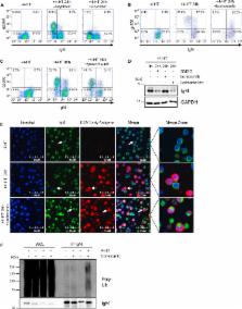
          Epstein-Barr virus (EBV) replication contributes to multiple human diseases, including infectious mononucleosis, nasopharyngeal carcinoma, B cell lymphomas, and oral hairy leukoplakia. We performed systematic quantitative analyses of temporal changes in host and EBV proteins during lytic replication to gain insights into virus-host interactions, using conditional Burkitt lymphoma models of type I and II EBV infection. We quantified profiles of >8,000 cellular and 69 EBV proteins, including >500 plasma membrane proteins, providing temporal views of the lytic B cell proteome and EBV virome. Our approach revealed EBV-induced remodeling of cell cycle, innate and adaptive immune pathways, including upregulation of the complement cascade and proteasomal degradation of the B cell receptor complex, conserved between EBV types I and II. Cross-comparison with proteomic analyses of human cytomegalovirus infection and of a Kaposi-sarcoma-associated herpesvirus immunoevasin identified host factors targeted by multiple herpesviruses. Our results provide an important resource for studies of EBV replication.

          Graphical Abstract

          Highlights

          • Unbiased global analysis of host and EBV proteome remodeling in lytic replication

          • Temporal profiles of >8,000 host and 69 viral proteins, using type I and II EBV

          • Both EBV types target the B cell receptor complex for degradation

          • Conserved EBV and HCMV lytic cycle host targets are identified

          Abstract

          Ersing et al. present a temporal proteomic map of EBV B cell lytic replication. Tandem-mass-tag-based proteomics uncover extensive remodeling of the human proteome by EBV, conserved across the two major EBV strains. Cell-cycle, innate, and adaptive immune pathways are modulated, complement is upregulated, and the B cell receptor is degraded by infection.

          Related collections

          Most cited references55

          • Record: found
          • Abstract: found
          • Article: not found

          IRF family of transcription factors as regulators of host defense.

          Interferon regulatory factors (IRFs) constitute a family of transcription factors that commonly possess a novel helix-turn-helix DNA-binding motif. Following the initial identification of two structurally related members, IRF-1 and IRF-2, seven additional members have now been reported. In addition, virally encoded IRFs, which may interfere with cellular IRFs, have also been identified. Thus far, intensive functional analyses have been done on IRF-1, revealing a remarkable functional diversity of this transcription factor in the regulation of cellular response in host defense. Indeed, IRF-1 selectively modulates different sets of genes, depending on the cell type and/or the nature of cellular stimuli, in order to evoke appropriate responses in each. More recently, much attention has also been focused on other IRF family members. Their functional roles, through interactions with their own or other members of the family of transcription factors, are becoming clearer in the regulation of host defense, such as innate and adaptive immune responses and oncogenesis.
            Bookmark
            • Record: found
            • Abstract: found
            • Article: not found

            A “Proteomic Ruler” for Protein Copy Number and Concentration Estimation without Spike-in Standards*

            Absolute protein quantification using mass spectrometry (MS)-based proteomics delivers protein concentrations or copy numbers per cell. Existing methodologies typically require a combination of isotope-labeled spike-in references, cell counting, and protein concentration measurements. Here we present a novel method that delivers similar quantitative results directly from deep eukaryotic proteome datasets without any additional experimental steps. We show that the MS signal of histones can be used as a “proteomic ruler” because it is proportional to the amount of DNA in the sample, which in turn depends on the number of cells. As a result, our proteomic ruler approach adds an absolute scale to the MS readout and allows estimation of the copy numbers of individual proteins per cell. We compare our protein quantifications with values derived via the use of stable isotope labeling by amino acids in cell culture and protein epitope signature tags in a method that combines spike-in protein fragment standards with precise isotope label quantification. The proteomic ruler approach yields quantitative readouts that are in remarkably good agreement with results from the precision method. We attribute this surprising result to the fact that the proteomic ruler approach omits error-prone steps such as cell counting or protein concentration measurements. The proteomic ruler approach is readily applicable to any deep eukaryotic proteome dataset—even in retrospective analysis—and we demonstrate its usefulness with a series of mouse organ proteomes.
              Bookmark
              • Record: found
              • Abstract: found
              • Article: not found

              Terminal differentiation into plasma cells initiates the replicative cycle of Epstein-Barr virus in vivo.

              In this paper we demonstrate that the cells which initiate replication of Epstein-Barr virus (EBV) in the tonsils of healthy carriers are plasma cells (CD38hi, CD10-, CD19+, CD20lo, surface immunoglobulin negative, and cytoplasmic immunoglobulin positive). We further conclude that differentiation into plasma cells, and not the signals that induce differentiation, initiates viral replication. This was confirmed by in vitro studies showing that the promoter for BZLF1, the gene that begins viral replication, becomes active only after memory cells differentiate into plasma cells and is also active in plasma cell lines. This differs from the reactivation of BZLF1 in vitro, which occurs acutely and is associated with apoptosis and not with differentiation. We suggest that differentiation and acute stress represent two distinct pathways of EBV reactivation in vivo. The fraction of cells replicating the virus decreases as the cells progress through the lytic cycle such that only a tiny fraction actually release infectious virus. This may reflect abortive replication or elimination of cells by the cellular immune response. Consistent with the later conclusion, the cells did not down regulate major histocompatibility complex class I molecules, suggesting that this is not an immune evasion tactic used by EBV and that the cells remain vulnerable to cytotoxic-T-lymphocyte attack.
                Bookmark

                Author and article information

                Contributors
                Journal
                Cell Rep
                Cell Rep
                Cell Reports
                Cell Press
                2211-1247
                16 May 2017
                16 May 2017
                16 May 2017
                : 19
                : 7
                : 1479-1493
                Affiliations
                [1 ]Division of Infectious Disease, Department of Medicine, Brigham & Women’s Hospital, Harvard Medical School, 181 Longwood Avenue, Boston, MA 02115, USA
                [2 ]Institut für Klinische und Molekulare Virologie, Friedrich-Alexander-Universität Erlangen-Nürnberg, 91054 Erlangen, Germany
                [3 ]Cambridge Institute for Medical Research, University of Cambridge, Hills Road, Cambridge CB2 0XY, UK
                [4 ]Department of Cell Biology, Harvard Medical School, 240 Longwood Avenue, Boston, MA 02115, USA
                [5 ]Wellcome Trust Sanger Institute, Hinxton, Cambridge, CB10 1HH, UK
                [6 ]Department of Immunobiology and Microbiology, Harvard Medical School, 77 Avenue Louis Pasteur, Boston, MA 02115, USA
                [7 ]Harvard Virology Program, Harvard Medical School, 77 Avenue Louis Pasteur, Boston, MA 02115, USA
                Author notes
                []Corresponding author mpw1001@ 123456cam.ac.uk
                [∗∗ ]Corresponding author bgewurz@ 123456partners.org
                [8]

                These authors contributed equally

                [9]

                These authors contributed equally

                [10]

                Lead Contact

                Article
                S2211-1247(17)30573-9
                10.1016/j.celrep.2017.04.062
                5446956
                28514666
                aa05f1cb-9b1c-47f8-bac6-4b39250c9d7e
                © 2017 The Author(s)

                This is an open access article under the CC BY license (http://creativecommons.org/licenses/by/4.0/).

                History
                : 22 December 2016
                : 24 March 2017
                : 20 April 2017
                Categories
                Resource

                Cell biology
                epstein-barr virus,herpesvirus,lytic replication,quantitative proteomics,tandem mass tag,host-pathogen interaction,immune evasion,b cell receptor,complement,viral evasion

                Comments

                Comment on this article