11
views
0
recommends
+1 Recommend
0 collections
    0
    shares
      • Record: found
      • Abstract: found
      • Article: found
      Is Open Access

      Toxicological Effects of Cadmium on Mammalian Testis

      review-article

      Read this article at

      Bookmark
          There is no author summary for this article yet. Authors can add summaries to their articles on ScienceOpen to make them more accessible to a non-specialist audience.

          Abstract

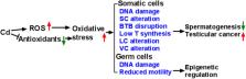
          Cadmium is a heavy metal, and people are exposed to it through contaminated foods and smoking. In humans and other mammals, cadmium causes damage to male testis. In this review, we summarize the effects of cadmium on the development and function of the testis. Cadmium causes severe structural damage to the seminiferous tubules, Sertoli cells, and blood-testis barrier, thus leading to the loss of sperm. Cadmium hinders Leydig cell development, inhibits Leydig cell function, and induces Leydig cell tumors. Cadmium also disrupts the vascular system of the testis. Cadmium is a reactive oxygen species inducer and possibly induces DNA damage, thus epigenetically regulating somatic cell and germ cell function, leading to male subfertility/infertility.

          Related collections

          Most cited references105

          • Record: found
          • Abstract: found
          • Article: not found

          Cadmium: toxic effects on the reproductive system and the embryo.

          The heavy metal cadmium (Cd) is a pollutant associated with several modern industrial processes. Cd is absorbed in significant quantities from cigarette smoke, and is known to have numerous undesirable effects on health in both experimental animals and humans, targeting the kidneys, liver and vascular systems in particular. However, a wide spectrum of deleterious effects on the reproductive tissues and the developing embryo has also been described. In the testis, changes due to disruption of the blood-testis barrier and oxidative stress have been noted, with onset of widespread necrosis at higher dosage exposures. Incorporation of Cd into the chromatin of the developing gamete has also been demonstrated. Ovarian Cd concentration increases with age, and has been associated with failure of progression of oocyte development from primary to secondary stage, and failure to ovulate. A further mechanism by which ovulation could be rendered ineffective is by failure of pick-up of the oocyte by the tubal cilia due to suboptimal expansion of the oocyte-cumulus complex and mis-expression of cell adhesion molecules. Retardation of trophoblastic outgrowth and development, placental necrosis and suppression of steroid biosynthesis, and altered handling of nutrient metals by the placenta all contribute to implantation delay and possible early pregnancy loss. Cd has been shown to accumulate in embryos from the four-cell stage onwards, and higher dosage exposure inhibits progression to the blastocyst stage, and can cause degeneration and decompaction in blastocysts following formation, with apoptosis and breakdown in cell adhesion. Following implantation, exposure of experimental animals to oral or parenteral Cd causes a wide range of abnormalities in the embryo, depending on the stage of exposure and dose given. Craniofacial, neurological, cardiovascular, gastrointestinal, genitourinary, and limb anomalies have all been described in placentates, with axial abnormalities and defects in somite structure noted in fish and ventral body wall defect and vertebral malformation occurring in the chick. In this paper, we examine the mechanisms by which Cd can affect reproductive health, and consider the use of micronutrients in prevention of these problems.
            Bookmark
            • Record: found
            • Abstract: found
            • Article: found
            Is Open Access

            The Dynamic Transcriptional Cell Atlas of Testis Development during Human Puberty

            Summary The human testis undergoes dramatic developmental and structural changes during puberty, including proliferation and maturation of somatic niche cells, and the onset of spermatogenesis. To characterize this understudied process, we profiled and analyzed single-cell transcriptomes of ∼10,000 testicular cells from four boys spanning puberty and compared them to those of infants and adults. During puberty, undifferentiated spermatogonia sequentially expand and differentiate prior to the initiation of gametogenesis. Notably, we identify a common pre-pubertal progenitor for Leydig and myoid cells and delineate candidate factors controlling pubertal differentiation. Furthermore, pre-pubertal Sertoli cells exhibit two distinct transcriptional states differing in metabolic profiles before converging to an alternative single mature population during puberty. Roles for testosterone in Sertoli cell maturation, antimicrobial peptide secretion, and spermatogonial differentiation are further highlighted through single-cell analysis of testosterone-suppressed transfemale testes. Taken together, our transcriptional atlas of the developing human testis provides multiple insights into developmental changes and key factors accompanying male puberty.
              Bookmark
              • Record: found
              • Abstract: found
              • Article: not found

              Pdgfr-alpha mediates testis cord organization and fetal Leydig cell development in the XY gonad.

              During testis development, the rapid morphological changes initiated by Sry require the coordinate integration of many signaling pathways. Based on the established role of the platelet-derived growth factor (PDGF) family of ligands and receptors in migration, proliferation, and differentiation of cells in various organ systems, we have investigated the role of PDGF in testis organogenesis. Analysis of expression patterns and characterization of the gonad phenotype in Pdgfr-alpha(-/-) embryos identified PDGFR-alpha as a critical mediator of signaling in the early testis at multiple steps of testis development. Pdgfr-alpha(-/-) XY gonads displayed disruptions in the organization of the vasculature and in the partitioning of interstitial and testis cord compartments. Closer examination revealed severe reductions in characteristic XY proliferation, mesonephric cell migration, and fetal Leydig cell differentiation. This work identifies PDGF signaling through the alpha receptor as an important event downstream of Sry in testis organogenesis and Leydig cell differentiation.
                Bookmark

                Author and article information

                Contributors
                Journal
                Front Genet
                Front Genet
                Front. Genet.
                Frontiers in Genetics
                Frontiers Media S.A.
                1664-8021
                26 May 2020
                2020
                : 11
                : 527
                Affiliations
                [1] 1Department of Obstetrics and Gynecology, The Second Affiliated Hospital and Yuying Children's Hospital of Wenzhou Medical University , Wenzhou, China
                [2] 2Department of Anesthesiology, The Second Affiliated Hospital and Yuying Children's Hospital of Wenzhou Medical University , Wenzhou, China
                Author notes

                Edited by: Md Saidur Rahman, Chung-Ang University, South Korea

                Reviewed by: Soterios A. Kyrtopoulos, National Hellenic Research Foundation, Greece; Richard John Brennan, Sanofi U. S., United States

                *Correspondence: Ren-Shan Ge r_ge@ 123456yahoo.com

                This article was submitted to Toxicogenomics, a section of the journal Frontiers in Genetics

                Article
                10.3389/fgene.2020.00527
                7265816
                32528534
                b8597bea-37c1-4e48-b061-89453766849e
                Copyright © 2020 Zhu, Li and Ge.

                This is an open-access article distributed under the terms of the Creative Commons Attribution License (CC BY). The use, distribution or reproduction in other forums is permitted, provided the original author(s) and the copyright owner(s) are credited and that the original publication in this journal is cited, in accordance with accepted academic practice. No use, distribution or reproduction is permitted which does not comply with these terms.

                History
                : 11 December 2019
                : 30 April 2020
                Page count
                Figures: 1, Tables: 1, Equations: 0, References: 114, Pages: 9, Words: 8619
                Funding
                Funded by: National Natural Science Foundation of China 10.13039/501100001809
                Funded by: Department of Health of Zhejiang Province 10.13039/501100008856
                Categories
                Genetics
                Review

                Genetics
                cadmium,reactive oxygen species,male infertility,spermatogenesis,leydig cell,testosterone
                Genetics
                cadmium, reactive oxygen species, male infertility, spermatogenesis, leydig cell, testosterone

                Comments

                Comment on this article