7
views
0
recommends
+1 Recommend
0 collections
    0
    shares
      • Record: found
      • Abstract: found
      • Article: found
      Is Open Access

      Double-edged-sword effect of IL-1 β on the osteogenesis of periodontal ligament stem cells via crosstalk between the NF- κB, MAPK and BMP/Smad signaling pathways

      research-article

      Read this article at

      Bookmark
          There is no author summary for this article yet. Authors can add summaries to their articles on ScienceOpen to make them more accessible to a non-specialist audience.

          Abstract

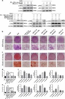
          Microenvironmental conditions can interfere with the functional role and differentiation of mesenchymal stem cells (MSCs). Recent studies suggest that an inflammatory microenvironment can significantly impact the osteogenic potential of periodontal ligament stem cells (PDLSCs), but the precise effects and mechanisms involved remain unclear. Here, we show for the first time that interleukin-1 β (IL-1 β) has dual roles in the osteogenesis of PDLSCs at concentrations ranging from physiologically healthy levels to those found in chronic periodontitis. Low doses of IL-1 β activate the BMP/Smad signaling pathway to promote the osteogenesis of PDLSCs, but higher doses of IL-1 β inhibit BMP/Smad signaling through the activation of nuclear factor- κB (NF- κB) and mitogen-activated protein kinase (MAPK) signaling, inhibiting osteogenesis. These results demonstrate that crosstalk between NF- κB, MAPK and BMP/Smad signaling mediates this dual effect of IL-1 β on PDLSCs. We also show that the impaired osteogenesis of PDLSCs results in more inflammatory cytokines and chemokines being released, inducing the chemotaxis of macrophages, which further clarifies the role of PDLSCs in the pathogenesis of periodontitis.

          Related collections

          Most cited references33

          • Record: found
          • Abstract: found
          • Article: found
          Is Open Access

          TGF-β and BMP Signaling in Osteoblast Differentiation and Bone Formation

          Transforming growth factor-beta (TGF-β)/bone morphogenic protein (BMP) signaling is involved in a vast majority of cellular processes and is fundamentally important throughout life. TGF-β/BMPs have widely recognized roles in bone formation during mammalian development and exhibit versatile regulatory functions in the body. Signaling transduction by TGF-β/BMPs is specifically through both canonical Smad-dependent pathways (TGF-β/BMP ligands, receptors and Smads) and non-canonical Smad-independent signaling pathway (e.g. p38 mitogen-activated protein kinase pathway, MAPK). Following TGF-β/BMP induction, both the Smad and p38 MAPK pathways converge at the Runx2 gene to control mesenchymal precursor cell differentiation. The coordinated activity of Runx2 and TGF-β/BMP-activated Smads is critical for formation of the skeleton. Recent advances in molecular and genetic studies using gene targeting in mice enable a better understanding of TGF-β/BMP signaling in bone and in the signaling networks underlying osteoblast differentiation and bone formation. This review summarizes the recent advances in our understanding of TGF-β/BMP signaling in bone from studies of genetic mouse models and human diseases caused by the disruption of TGF-β/BMP signaling. This review also highlights the different modes of cross-talk between TGF-β/BMP signaling and the signaling pathways of MAPK, Wnt, Hedgehog, Notch, and FGF in osteoblast differentiation and bone formation.
            Bookmark
            • Record: found
            • Abstract: found
            • Article: not found

            Mesenchymal stem cells derived from dental tissues vs. those from other sources: their biology and role in regenerative medicine.

            To date, 5 different human dental stem/progenitor cells have been isolated and characterized: dental pulp stem cells (DPSCs), stem cells from exfoliated deciduous teeth (SHED), periodontal ligament stem cells (PDLSCs), stem cells from apical papilla (SCAP), and dental follicle progenitor cells (DFPCs). These postnatal populations have mesenchymal-stem-cell-like (MSC) qualities, including the capacity for self-renewal and multilineage differentiation potential. MSCs derived from bone marrow (BMMSCs) are capable of giving rise to various lineages of cells, such as osteogenic, chondrogenic, adipogenic, myogenic, and neurogenic cells. The dental-tissue-derived stem cells are isolated from specialized tissue with potent capacities to differentiate into odontogenic cells. However, they also have the ability to give rise to other cell lineages similar to, but different in potency from, that of BMMSCs. This article will review the isolation and characterization of the properties of different dental MSC-like populations in comparison with those of other MSCs, such as BMMSCs. Important issues in stem cell biology, such as stem cell niche, homing, and immunoregulation, will also be discussed.
              Bookmark
              • Record: found
              • Abstract: found
              • Article: not found

              Phosphorylation of NF-kappaB and IkappaB proteins: implications in cancer and inflammation.

              Nuclear factor-kappaB (NF-kappaB) is a transcription factor that has crucial roles in inflammation, immunity, cell proliferation and apoptosis. Activation of NF-kappaB mainly occurs via IkappaB kinase (IKK)-mediated phosphorylation of inhibitory molecules, including IkappaBalpha. Optimal induction of NF-kappaB target genes also requires phosphorylation of NF-kappaB proteins, such as p65, within their transactivation domain by a variety of kinases in response to distinct stimuli. Whether, and how, phosphorylation modulates the function of other NF-kappaB and IkappaB proteins, such as B-cell lymphoma 3, remains unclear. The identification and characterization of all the kinases known to phosphorylate NF-kappaB and IkappaB proteins are described here. Because deregulation of NF-kappaB and IkappaB phosphorylations is a hallmark of chronic inflammatory diseases and cancer, newly designed drugs targeting these constitutively activated signalling pathways represent promising therapeutic tools.
                Bookmark

                Author and article information

                Journal
                Cell Death Dis
                Cell Death Dis
                Cell Death & Disease
                Nature Publishing Group
                2041-4889
                July 2016
                14 July 2016
                1 July 2016
                : 7
                : 7
                : e2296
                Affiliations
                [1 ]Department of Prosthodontics, Shanghai Key Laboratory of Stomatology, Shanghai Ninth People's Hospital, Shanghai JiaoTong University School of Medicine , 639 Zhizaoju Road, Shanghai, China
                [2 ]Shanghai Key Laboratory of Orthopaedic Implant, Department of Orthopaedics Surgery, Shanghai Ninth People's Hospital, Shanghai JiaoTong University School of Medicine , 639 Zhizaoju Road, Shanghai, China
                Author notes
                [* ]Shanghai Key Laboratory of Orthopaedic Implant, Department of Orthopaedics Surgery, Shanghai Ninth People's Hospital, Shanghai JiaoTong University School of Medicine , 639 Zhizaoju Road, Shanghai 200011, China. Tel/Fax: +86 21 6313 7020; E-mail: tingtingtang@ 123456hotmail.com
                [* ]Department of Prosthodontics, Shanghai Key Laboratory of Stomatology, Shanghai Ninth People's Hospital, Shanghai JiaoTong University School of Medicine , 639 Zhizaoju Road, Shanghai 200011, China. Tel/Fax: +86 21 6308 7768, E-mail: lueryi222@ 123456126.com
                Article
                cddis2016204
                10.1038/cddis.2016.204
                4973347
                27415426
                ba77b5ca-08ec-48b0-a2a7-884c4315aea0
                Copyright © 2016 The Author(s)

                Cell Death and Disease is an open-access journal published by Nature Publishing Group. This work is licensed under a Creative Commons Attribution 4.0 International License. The images or other third party material in this article are included in the article's Creative Commons license, unless indicated otherwise in the credit line; if the material is not included under the Creative Commons license, users will need to obtain permission from the license holder to reproduce the material. To view a copy of this license, visit http://creativecommons.org/licenses/by/4.0/

                History
                : 21 March 2016
                : 11 June 2016
                : 14 June 2016
                Categories
                Original Article

                Cell biology
                Cell biology

                Comments

                Comment on this article