8
views
0
recommends
+1 Recommend
0 collections
    0
    shares
      • Record: found
      • Abstract: found
      • Article: found
      Is Open Access

      Inactivation Kinetics and Mechanical Gating of Piezo1 Ion Channels Depend on Subdomains within the Cap

      research-article
      1 , 1 , 2 , *
      Cell reports

      Read this article at

      Bookmark
          There is no author summary for this article yet. Authors can add summaries to their articles on ScienceOpen to make them more accessible to a non-specialist audience.

          SUMMARY

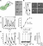
          Piezo1 ion channels are activated by mechanical stimuli and mediate the sensing of blood flow. Although cryo-electron microscopy (cryo-EM) structures have revealed the overall architecture of Piezo1, the precise domains involved in activation and subsequent inactivation have remained elusive. Here, we perform a targeted chimeric screen between Piezo1 and the closely related isoform Piezo2 and use electrophysiology to characterize their inactivation kinetics during mechanical stimulation. We identify three small subdomains within the extracellular cap that individually can confer the distinct kinetics of inactivation of Piezo2 onto Piezo1. We further show by cysteine crosslinking that conformational flexibility of these subdomains is required for mechanical activation to occur and that electrostatic interactions functionally couple the cap to the extensive blades, which have been proposed to function as sensors of membrane curvature and tension. This study provides a demonstration of internal gating motions involved in mechanotransduction by Piezo1.

          Graphical Abstract

          In Brief

          Lewis and Grandl combine a chimeric screen and cysteine crosslinking to identify small subdomains of the cap of mechanically activated Piezo1 ion channels that must have conformational flexibility for mechanical gating. They further show that electrostatic interactions couple one of these domains to the channel blade.

          Related collections

          Most cited references36

          • Record: found
          • Abstract: found
          • Article: not found

          Piezos are pore-forming subunits of mechanically activated channels

          Mechanotransduction plays a crucial role in physiology. Biological processes including sensing touch and sound waves require yet unidentified cation channels that detect pressure. Mouse piezo1 (mpiezo1) and mpiezo2 induce mechanically activated cationic currents in cells; however, it is unknown if piezos are pore-forming ion channels or modulate ion channels. We show that Drosophila piezo (dpiezo) also induces mechanically activated currents in cells, but through channels with remarkably distinct pore properties including sensitivity to the pore blocker ruthenium red and single channel conductances. mpiezo1 assembles as a ~1.2 million-Dalton tetramer, with no evidence of other proteins in this complex. Finally, purified mpiezo1 reconstituted into asymmetric lipid bilayers and liposomes forms ruthenium red-sensitive ion channels. These data demonstrate that piezos are an evolutionarily conserved ion channel family involved in mechanotransduction.
            Bookmark
            • Record: found
            • Abstract: found
            • Article: not found

            Stretch-activated ion channel Piezo1 directs lineage choice in human neural stem cells.

            Neural stem cells are multipotent cells with the ability to differentiate into neurons, astrocytes, and oligodendrocytes. Lineage specification is strongly sensitive to the mechanical properties of the cellular environment. However, molecular pathways transducing matrix mechanical cues to intracellular signaling pathways linked to lineage specification remain unclear. We found that the mechanically gated ion channel Piezo1 is expressed by brain-derived human neural stem/progenitor cells and is responsible for a mechanically induced ionic current. Piezo1 activity triggered by traction forces elicited influx of Ca(2+), a known modulator of differentiation, in a substrate-stiffness-dependent manner. Inhibition of channel activity by the pharmacological inhibitor GsMTx-4 or by siRNA-mediated Piezo1 knockdown suppressed neurogenesis and enhanced astrogenesis. Piezo1 knockdown also reduced the nuclear localization of the mechanoreactive transcriptional coactivator Yes-associated protein. We propose that the mechanically gated ion channel Piezo1 is an important determinant of mechanosensitive lineage choice in neural stem cells and may play similar roles in other multipotent stem cells.
              Bookmark
              • Record: found
              • Abstract: found
              • Article: found
              Is Open Access

              Piezo1 links mechanical forces to red blood cell volume

              Red blood cells (RBCs) experience significant mechanical forces while recirculating, but the consequences of these forces are not fully understood. Recent work has shown that gain-of-function mutations in mechanically activated Piezo1 cation channels are associated with the dehydrating RBC disease xerocytosis, implicating a role of mechanotransduction in RBC volume regulation. However, the mechanisms by which these mutations result in RBC dehydration are unknown. In this study, we show that RBCs exhibit robust calcium entry in response to mechanical stretch and that this entry is dependent on Piezo1 expression. Furthermore, RBCs from blood-cell-specific Piezo1 conditional knockout mice are overhydrated and exhibit increased fragility both in vitro and in vivo. Finally, we show that Yoda1, a chemical activator of Piezo1, causes calcium influx and subsequent dehydration of RBCs via downstream activation of the KCa3.1 Gardos channel, directly implicating Piezo1 signaling in RBC volume control. Therefore, mechanically activated Piezo1 plays an essential role in RBC volume homeostasis. DOI: http://dx.doi.org/10.7554/eLife.07370.001
                Bookmark

                Author and article information

                Journal
                101573691
                39703
                Cell Rep
                Cell Rep
                Cell reports
                2211-1247
                31 January 2020
                21 January 2020
                15 February 2020
                : 30
                : 3
                : 870-880.e2
                Affiliations
                [1 ]Department of Neurobiology, Duke University Medical Center, Durham, NC 27710, USA
                [2 ]Lead Contact
                Author notes

                AUTHOR CONTRIBUTIONS

                A.H.L. performed all experiments and data analysis. A.H.L. and J.G. designed the study and wrote the manuscript.

                [* ]Correspondence: grandl@ 123456neuro.duke.edu
                Article
                NIHMS1551547
                10.1016/j.celrep.2019.12.040
                7021530
                31968259
                ba97b090-68b2-40a1-8ec3-4653269d4ec0

                This is an open access article under the CC BY-NC-ND license ( http://creativecommons.org/licenses/by-nc-nd/4.0/).

                History
                Categories
                Article

                Cell biology
                Cell biology

                Comments

                Comment on this article