4
views
0
recommends
+1 Recommend
0 collections
    0
    shares
      • Record: found
      • Abstract: found
      • Article: found
      Is Open Access

      Unravelling HP1 functions: post-transcriptional regulation of stem cell fate

      review-article

      Read this article at

      Bookmark
          There is no author summary for this article yet. Authors can add summaries to their articles on ScienceOpen to make them more accessible to a non-specialist audience.

          Abstract

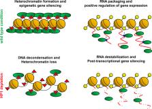
          Heterochromatin protein 1 (HP1) is a non-histone chromosomal protein first identified in Drosophila as a major component of constitutive heterochromatin, required for stable epigenetic gene silencing in many species including humans. Over the years, several studies have highlighted additional roles of HP1 in different cellular processes including telomere maintenance, DNA replication and repair, chromosome segregation and, surprisingly, positive regulation of gene expression. In this review, we briefly summarize past research and recent results supporting the unexpected and emerging role of HP1 in activating gene expression. In particular, we discuss the role of HP1 in post-transcriptional regulation of mRNA processing because it has proved decisive in the control of germline stem cells homeostasis in Drosophila and has certainly added a new dimension to our understanding on HP1 targeting and functions in epigenetic regulation of stem cell behaviour.

          Related collections

          Most cited references110

          • Record: found
          • Abstract: found
          • Article: not found

          Selective recognition of methylated lysine 9 on histone H3 by the HP1 chromo domain.

          Heterochromatin protein 1 (HP1) is localized at heterochromatin sites where it mediates gene silencing. The chromo domain of HP1 is necessary for both targeting and transcriptional repression. In the fission yeast Schizosaccharomyces pombe, the correct localization of Swi6 (the HP1 equivalent) depends on Clr4, a homologue of the mammalian SUV39H1 histone methylase. Both Clr4 and SUV39H1 methylate specifically lysine 9 of histone H3 (ref. 6). Here we show that HP1 can bind with high affinity to histone H3 methylated at lysine 9 but not at lysine 4. The chromo domain of HP1 is identified as its methyl-lysine-binding domain. A point mutation in the chromo domain, which destroys the gene silencing activity of HP1 in Drosophila, abolishes methyl-lysine-binding activity. Genetic and biochemical analysis in S. pombe shows that the methylase activity of Clr4 is necessary for the correct localization of Swi6 at centromeric heterochromatin and for gene silencing. These results provide a stepwise model for the formation of a transcriptionally silent heterochromatin: SUV39H1 places a 'methyl marker' on histone H3, which is then recognized by HP1 through its chromo domain. This model may also explain the stable inheritance of the heterochromatic state.
            Bookmark
            • Record: found
            • Abstract: found
            • Article: not found

            Phase separation drives heterochromatin domain formation

            Constitutive heterochromatin is an important component of eukaryotic genomes that has essential roles in nuclear architecture, DNA repair and genome stability, and silencing of transposon and gene expression. Heterochromatin is highly enriched for repetitive sequences, and is defined epigenetically by methylation of histone H3 at lysine 9 and recruitment of its binding partner heterochromatin protein 1 (HP1). A prevalent view of heterochromatic silencing is that these and associated factors lead to chromatin compaction, resulting in steric exclusion of regulatory proteins such as RNA polymerase from the underlying DNA. However, compaction alone does not account for the formation of distinct, multi-chromosomal, membrane-less heterochromatin domains within the nucleus, fast diffusion of proteins inside the domain, and other dynamic features of heterochromatin. Here we present data that support an alternative hypothesis: that the formation of heterochromatin domains is mediated by phase separation, a phenomenon that gives rise to diverse non-membrane-bound nuclear, cytoplasmic and extracellular compartments. We show that Drosophila HP1a protein undergoes liquid–liquid demixing in vitro, and nucleates into foci that display liquid properties during the first stages of heterochromatin domain formation in early Drosophila embryos. Furthermore, in both Drosophila and mammalian cells, heterochromatin domains exhibit dynamics that are characteristic of liquid phase-separation, including sensitivity to the disruption of weak hydrophobic interactions, and reduced diffusion, increased coordinated movement and inert probe exclusion at the domain boundary. We conclude that heterochromatic domains form via phase separation, and mature into a structure that includes liquid and stable compartments. We propose that emergent biophysical properties associated with phase-separated systems are critical to understanding the unusual behaviours of heterochromatin, and how chromatin domains in general regulate essential nuclear functions.
              Bookmark
              • Record: found
              • Abstract: found
              • Article: not found

              Liquid droplet formation by HP1α suggests a role for phase separation in heterochromatin

              Gene silencing by heterochromatin is proposed to occur in part from the ability of HP1 proteins to spread across large regions of the genome, compact the underlying chromatin and recruit repressive activities 1–3 . Here we identify a new property of the human HP1α protein: the ability to form phase-separated droplets. While unmodified HP1α is soluble, either phosphorylation of its N-terminal extension or DNA binding promotes the formation of phase-separated droplets. Phosphorylation driven phase-separation can be promoted or reversed by specific HP1α ligands. Known components of heterochromatin such as nucleosomes and DNA preferentially partition into the HP1α droplets but other molecules such as the transcription factor TFIIB show no preference. Using single-molecule DNA curtains we find that unmodified and phosphorylated HP1α induce rapid compaction of DNA strands into puncta, though with different characteristics. We show by direct protein delivery into mammalian cells that an HP1α mutant incapable of phase separation in vitro forms smaller and fewer nuclear puncta than phosphorylated HP1α. These findings suggest that heterochromatin mediated gene silencing may occur in part through sequestration of compacted chromatin in phase-separated HP1 droplets, which are dissolved or formed by specific ligands based on nuclear context.
                Bookmark

                Author and article information

                Contributors
                assuntamaria.casale@uniroma1.it
                lucia.piacentini@uniroma1.it
                Journal
                Chromosoma
                Chromosoma
                Chromosoma
                Springer Berlin Heidelberg (Berlin/Heidelberg )
                0009-5915
                1432-0886
                15 June 2021
                15 June 2021
                2021
                : 130
                : 2-3
                : 103-111
                Affiliations
                GRID grid.7841.a, Department of Biology and Biotechnology “C. Darwin”, , Sapienza University of Rome, ; Rome, Italy
                Author information
                http://orcid.org/0000-0002-7025-7368
                http://orcid.org/0000-0002-5862-1390
                http://orcid.org/0000-0002-3656-5068
                Article
                760
                10.1007/s00412-021-00760-1
                8426308
                34128099
                dcba45b4-0e2c-4f21-b7e4-b20f063fa47b
                © The Author(s) 2021

                Open AccessThis article is licensed under a Creative Commons Attribution 4.0 International License, which permits use, sharing, adaptation, distribution and reproduction in any medium or format, as long as you give appropriate credit to the original author(s) and the source, provide a link to the Creative Commons licence, and indicate if changes were made. The images or other third party material in this article are included in the article's Creative Commons licence, unless indicated otherwise in a credit line to the material. If material is not included in the article's Creative Commons licence and your intended use is not permitted by statutory regulation or exceeds the permitted use, you will need to obtain permission directly from the copyright holder. To view a copy of this licence, visit http://creativecommons.org/licenses/by/4.0/.

                History
                : 4 February 2021
                : 17 May 2021
                : 1 June 2021
                Funding
                Funded by: Università degli Studi di Roma La Sapienza
                Categories
                Mini-Review
                Custom metadata
                © Springer-Verlag GmbH Germany, part of Springer Nature 2021

                Genetics
                heterochromatin protein 1,heterochromatin,post-transcriptional regulation of gene expression,germline stem cells,drosophila melanogaster

                Comments

                Comment on this article