156
views
0
recommends
+1 Recommend
0 collections
    0
    shares
      • Record: found
      • Abstract: found
      • Article: found
      Is Open Access

      Uniform nomenclature for the mitochondrial contact site and cristae organizing system

      review-article

      Read this article at

      Bookmark
          There is no author summary for this article yet. Authors can add summaries to their articles on ScienceOpen to make them more accessible to a non-specialist audience.

          Abstract

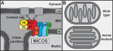
          The mitochondrial inner membrane contains a large protein complex that functions in inner membrane organization and formation of membrane contact sites. The complex was variably named the mitochondrial contact site complex, mitochondrial inner membrane organizing system, mitochondrial organizing structure, or Mitofilin/Fcj1 complex. To facilitate future studies, we propose to unify the nomenclature and term the complex “mitochondrial contact site and cristae organizing system” and its subunits Mic10 to Mic60.

          Related collections

          Most cited references37

          • Record: found
          • Abstract: found
          • Article: not found

          Translocation of proteins into mitochondria.

          About 10% to 15% of the nuclear genes of eukaryotic organisms encode mitochondrial proteins. These proteins are synthesized in the cytosol and recognized by receptors on the surface of mitochondria. Translocases in the outer and inner membrane of mitochondria mediate the import and intramitochondrial sorting of these proteins; ATP and the membrane potential are used as energy sources. Chaperones and auxiliary factors assist in the folding and assembly of mitochondrial proteins into their native, three-dimensional structures. This review summarizes the present knowledge on the import and sorting of mitochondrial precursor proteins, with a special emphasis on unresolved questions and topics of current research.
            Bookmark
            • Record: found
            • Abstract: found
            • Article: not found

            Macromolecular organization of ATP synthase and complex I in whole mitochondria.

            We used electron cryotomography to study the molecular arrangement of large respiratory chain complexes in mitochondria from bovine heart, potato, and three types of fungi. Long rows of ATP synthase dimers were observed in intact mitochondria and cristae membrane fragments of all species that were examined. The dimer rows were found exclusively on tightly curved cristae edges. The distance between dimers along the rows varied, but within the dimer the distance between F(1) heads was constant. The angle between monomers in the dimer was 70° or above. Complex I appeared as L-shaped densities in tomograms of reconstituted proteoliposomes. Similar densities were observed in flat membrane regions of mitochondrial membranes from all species except Saccharomyces cerevisiae and identified as complex I by quantum-dot labeling. The arrangement of respiratory chain proton pumps on flat cristae membranes and ATP synthase dimer rows along cristae edges was conserved in all species investigated. We propose that the supramolecular organization of respiratory chain complexes as proton sources and ATP synthase rows as proton sinks in the mitochondrial cristae ensures optimal conditions for efficient ATP synthesis.
              Bookmark
              • Record: found
              • Abstract: found
              • Article: not found

              The mitochondrial contact site complex, a determinant of mitochondrial architecture.

              Mitochondria are organelles with a complex architecture. They are bounded by an envelope consisting of the outer membrane and the inner boundary membrane (IBM). Narrow crista junctions (CJs) link the IBM to the cristae. OMs and IBMs are firmly connected by contact sites (CS). The molecular nature of the CS remained unknown. Using quantitative high-resolution mass spectrometry we identified a novel complex, the mitochondrial contact site (MICOS) complex, formed by a set of mitochondrial membrane proteins that is essential for the formation of CS. MICOS is preferentially located at the CJs. Upon loss of one of the MICOS subunits, CJs disappear completely or are impaired, showing that CJs require the presence of CS to form a superstructure that links the IBM to the cristae. Loss of MICOS subunits results in loss of respiratory competence and altered inheritance of mitochondrial DNA.
                Bookmark

                Author and article information

                Journal
                J Cell Biol
                J. Cell Biol
                jcb
                jcb
                The Journal of Cell Biology
                The Rockefeller University Press
                0021-9525
                1540-8140
                31 March 2014
                : 204
                : 7
                : 1083-1086
                Affiliations
                [1 ]Institut für Biochemie und Molekularbiologie, Zentrum für Biochemie und Molekulare Zellforschung , and [2 ]BIOSS Centre for Biological Signalling Studies, Universität Freiburg, 79104 Freiburg, Germany
                [3 ]Pasteur Institute of Rome, University of Rome at Sapienza, 00161 Rome, Italy
                [4 ]Metabolic Profiling, Inc., Eugene, OR 97401
                [5 ]Donnelly Centre for Cellular and Biomolecular Research and [6 ]Department of Molecular Genetics, University of Toronto, Toronto, Ontario M5S 3E1, Canada
                [7 ]The International Institute of Molecular and Cell Biology, 02-109 Warsaw, Poland
                [8 ]Howard Hughes Medical Institute , [9 ]Department of Pharmacology , and [10 ]Department of Chemistry and Biochemistry, University of California, San Diego, La Jolla, CA 92093
                [11 ]Department of Biochemistry II, University of Göttingen, 37073 Göttingen, Germany
                [12 ]Department of Biochemistry, University of Washington, Seattle, WA 98195
                [13 ]Somechi Orchid Laboratory, Chofu, Tokyo 182-0023, Japan
                [14 ]Department of Neurology, University Medical Center, 37075 Göttingen, Germany
                [15 ]Max Planck Institute for Biophysical Chemistry, 37077 Göttingen, Germany
                [16 ]The National Laboratory of Protein Engineering and Plant Genetic Engineering, College of Life Sciences, Peking University, Beijing, P.R. China 100871
                [17 ]Department of Microbiology, Biocenter, University of Würzburg, 97074 Würzburg, Germany
                [18 ]Department of Cell and Developmental Biology, University of Massachusetts Medical School, Worcester, MA 01655
                [19 ]Department of Life Sciences, Pohang University of Science and Technology, Pohang 790-784, South Korea
                [20 ]Mitochondrial Biology, Buchmann Institute for Molecular Life Sciences and [21 ]Centre for Molecular Medicine, Goethe University, 60438 Frankfurt am Main, Germany
                [22 ]Department of Pharmacology, State University of New York Upstate Medical University, Syracuse, NY 13210
                [23 ]Department of Molecular Cellular Oncology and Microbiology, Tokyo Medical and Dental University, Bunkyo-ku, Tokyo 113-8549, Japan
                [24 ]Department of Biological Chemistry, David Geffen School of Medicine, University of California, Los Angeles, Los Angeles, CA 90095
                [25 ]Molecular Cell Biology, University of Groningen, 9700 CC Groningen, Netherlands
                [26 ]Howard Hughes Medical Institute and [27 ]Department of Cellular and Molecular Pharmacology, University of California, San Francisco, San Francisco, CA 94158
                [28 ]Zellbiologie, Universität Bayreuth, 95440 Bayreuth, Germany
                [29 ]Crown Bioscience, Inc., Taicang City, Jiangsu Province, P.R. China 215400
                [30 ]Abteilung für Zelluläre Biochemie, Max-Planck-Institut für Biochemie, 82152 Martinsried, Germany
                [31 ]Department of Molecular and Cellular Biology, University of California, Davis, Davis, CA 95616
                Author notes
                Article
                201401006
                10.1083/jcb.201401006
                3971754
                24687277
                e4c4f949-f5f1-4218-a6bf-29cecbe9187e
                © 2014 Pfanner et al.

                This article is distributed under the terms of an Attribution–Noncommercial–Share Alike–No Mirror Sites license for the first six months after the publication date (see http://www.rupress.org/terms). After six months it is available under a Creative Commons License (Attribution–Noncommercial–Share Alike 3.0 Unported license, as described at http://creativecommons.org/licenses/by-nc-sa/3.0/).

                History
                : 2 January 2014
                : 6 March 2014
                Categories
                Reviews
                Comment

                Cell biology
                Cell biology

                Comments

                Comment on this article