26
views
0
recommends
+1 Recommend
0 collections
    0
    shares
      • Record: found
      • Abstract: found
      • Article: found
      Is Open Access

      A Pixel-Encoder Retinal Ganglion Cell with Spatially Offset Excitatory and Inhibitory Receptive Fields

      research-article
      1 , 2 , 1 , 1 , 3 , 4 , 5
      Cell reports

      Read this article at

      Bookmark
          There is no author summary for this article yet. Authors can add summaries to their articles on ScienceOpen to make them more accessible to a non-specialist audience.

          SUMMARY

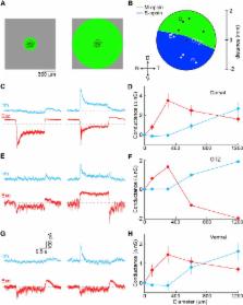
          The spike trains of retinal ganglion cells (RGCs) are the only source of visual information to the brain. Here, we genetically identify an RGC type in mice that functions as a pixel encoder and increases firing to light increments (Pix ON-RGC). Pix ON-RGCs have medium-sized dendritic arbors and non-canonical center-surround receptive fields. From their receptive field center, Pix ON-RGCs receive only excitatory input, which encodes contrast and spatial information linearly. From their receptive field surround, Pix ON-RGCs receive only inhibitory input, which is temporally matched to the excitatory center input. As a result, the firing rate of Pix ON-RGCs linearly encodes local image contrast. Spatially offset (i.e., truly lateral) inhibition of Pix ON-RGCs arises from spiking GABAergic amacrine cells. The receptive field organization of Pix ON-RGCs is independent of stimulus wavelength (i.e., achromatic). Pix ON-RGCs project predominantly to the dorsal lateral geniculate nucleus (dLGN) of the thalamus and likely contribute to visual perception.

          In Brief

          Johnson et al. genetically identify a pixel-encoder retinal ganglion cell type in mice (Pix ON-RGCs). Pix ON-RGCs have spatially offset excitatory and inhibitory receptive fields and encode local image contrast approximately linearly. Their axons project to the dorsolateral geniculate nucleus of the thalamus indicating that Pix ON-RGCs likely contribute to visual perception.

          Related collections

          Most cited references54

          • Record: found
          • Abstract: found
          • Article: not found

          How inhibition shapes cortical activity.

          Cortical processing reflects the interplay of synaptic excitation and synaptic inhibition. Rapidly accumulating evidence is highlighting the crucial role of inhibition in shaping spontaneous and sensory-evoked cortical activity and thus underscores how a better knowledge of inhibitory circuits is necessary for our understanding of cortical function. We discuss current views of how inhibition regulates the function of cortical neurons and point to a number of important open questions. Copyright © 2011 Elsevier Inc. All rights reserved.
            Bookmark
            • Record: found
            • Abstract: found
            • Article: not found

            The types of retinal ganglion cells: current status and implications for neuronal classification.

            In the retina, photoreceptors pass visual information to interneurons, which process it and pass it to retinal ganglion cells (RGCs). Axons of RGCs then travel through the optic nerve, telling the rest of the brain all it will ever know about the visual world. Research over the past several decades has made clear that most RGCs are not merely light detectors, but rather feature detectors, which send a diverse set of parallel, highly processed images of the world on to higher centers. Here, we review progress in classification of RGCs by physiological, morphological, and molecular criteria, making a particular effort to distinguish those cell types that are definitive from those for which information is partial. We focus on the mouse, in which molecular and genetic methods are most advanced. We argue that there are around 30 RGC types and that we can now account for well over half of all RGCs. We also use RGCs to examine the general problem of neuronal classification, arguing that insights and methods from the retina can guide the classification enterprise in other brain regions.
              Bookmark
              • Record: found
              • Abstract: not found
              • Article: not found

              Neuronal morphometry directly from bitmap images.

                Bookmark

                Author and article information

                Journal
                101573691
                39703
                Cell Rep
                Cell Rep
                Cell reports
                2211-1247
                15 February 2018
                06 February 2018
                26 February 2018
                : 22
                : 6
                : 1462-1472
                Affiliations
                [1 ]Department of Ophthalmology and Visual Sciences, Washington University School of Medicine, Saint Louis, MO 63110, USA
                [2 ]Graduate Program in Neuroscience, Washington University School of Medicine, Saint Louis, MO 63110, USA
                [3 ]Department of Neuroscience, Washington University School of Medicine, Saint Louis, MO 63110, USA
                [4 ]Department of Biomedical Engineering, Washington University School of Medicine, Saint Louis, MO 63110, USA
                [5 ]Hope Center for Neurological Disorders, Washington University School of Medicine, Saint Louis, MO 63110, USA
                Author notes
                [6]

                Lead Contact

                Article
                NIHMS943489
                10.1016/j.celrep.2018.01.037
                5826572
                29425502
                e714fb1b-de96-4762-bd49-4af958495bcf

                This is an open access article under the CC BY-NC-ND license ( http://creativecommons.org/licenses/by-nc-nd/4.0/).

                History
                Categories
                Article

                Cell biology
                Cell biology

                Comments

                Comment on this article