17
views
0
recommends
+1 Recommend
0 collections
    0
    shares
      • Record: found
      • Abstract: found
      • Article: found
      Is Open Access

      No ATG8s, no problem? How LC3/GABARAP proteins contribute to autophagy

      article-commentary
      The Journal of Cell Biology
      The Rockefeller University Press

      Read this article at

      Bookmark
          There is no author summary for this article yet. Authors can add summaries to their articles on ScienceOpen to make them more accessible to a non-specialist audience.

          Abstract

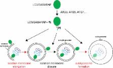
          Martens previews work by Nguyen et al. analyzing the essential functions of ATG8 family proteins LC3/GABARAPs in autophagy.

          Abstract

          The ATG8 family LC3/GABARAP proteins are attached to the membrane of nascent autophagosomes, but their functions during autophagy are unclear. In this issue, Nguyen et al. (2016. J. Cell Biol. https://doi.org/10.1083/jcb.201607039) show that LC3/GABARAP proteins are not essential for autophagosome formation but are critical for autophagosome–lysosome fusion.

          Related collections

          Most cited references8

          • Record: found
          • Abstract: found
          • Article: not found

          A ubiquitin-like system mediates protein lipidation.

          Autophagy is a dynamic membrane phenomenon for bulk protein degradation in the lysosome/vacuole. Apg8/Aut7 is an essential factor for autophagy in yeast. We previously found that the carboxy-terminal arginine of nascent Apg8 is removed by Apg4/Aut2 protease, leaving a glycine residue at the C terminus. Apg8 is then converted to a form (Apg8-X) that is tightly bound to the membrane. Here we report a new mode of protein lipidation. Apg8 is covalently conjugated to phosphatidylethanolamine through an amide bond between the C-terminal glycine and the amino group of phosphatidylethanolamine. This lipidation is mediated by a ubiquitination-like system. Apg8 is a ubiquitin-like protein that is activated by an E1 protein, Apg7 (refs 7, 8), and is transferred subsequently to the E2 enzymes Apg3/Aut1 (ref. 9). Apg7 activates two different ubiquitin-like proteins, Apg12 (ref. 10) and Apg8, and assigns them to specific E2 enzymes, Apg10 (ref. 11) and Apg3, respectively. These reactions are necessary for the formation of Apg8-phosphatidylethanolamine. This lipidation has an essential role in membrane dynamics during autophagy.
            Bookmark
            • Record: found
            • Abstract: found
            • Article: not found

            Discovery of Atg5/Atg7-independent alternative macroautophagy.

            Macroautophagy is a process that leads to the bulk degradation of subcellular constituents by producing autophagosomes/autolysosomes. It is believed that Atg5 (ref. 4) and Atg7 (ref. 5) are essential genes for mammalian macroautophagy. Here we show, however, that mouse cells lacking Atg5 or Atg7 can still form autophagosomes/autolysosomes and perform autophagy-mediated protein degradation when subjected to certain stressors. Although lipidation of the microtubule-associated protein light chain 3 (LC3, also known as Map1lc3a) to form LC3-II is generally considered to be a good indicator of macroautophagy, it did not occur during the Atg5/Atg7-independent alternative process of macroautophagy. We also found that this alternative process of macroautophagy was regulated by several autophagic proteins, including Unc-51-like kinase 1 (Ulk1) and beclin 1. Unlike conventional macroautophagy, autophagosomes seemed to be generated in a Rab9-dependent manner by the fusion of isolation membranes with vesicles derived from the trans-Golgi and late endosomes. In vivo, Atg5-independent alternative macroautophagy was detected in several embryonic tissues. It also had a function in clearing mitochondria during erythroid maturation. These results indicate that mammalian macroautophagy can occur through at least two different pathways: an Atg5/Atg7-dependent conventional pathway and an Atg5/Atg7-independent alternative pathway.
              Bookmark
              • Record: found
              • Abstract: found
              • Article: found
              Is Open Access

              Atg8 family LC3/GABARAP proteins are crucial for autophagosome–lysosome fusion but not autophagosome formation during PINK1/Parkin mitophagy and starvation

              Current autophagy models suggest that Atg8 family LC3/GABARAP proteins are essential mediators of autophagosome biogenesis. Nguyen et al. exploit CRISPR/Cas9-generated knockouts of the LC3 or GABARAP subfamilies, or both subfamilies, to show that Atg8s are dispensable for autophagosome biogenesis but essential for autophagosome–lysosome fusion.
                Bookmark

                Author and article information

                Journal
                J Cell Biol
                J. Cell Biol
                jcb
                jcb
                The Journal of Cell Biology
                The Rockefeller University Press
                0021-9525
                1540-8140
                19 December 2016
                : 215
                : 6
                : 761-763
                Affiliations
                [1]Department of Biochemistry and Cell Biology, Max F. Perutz Laboratories, University of Vienna, 1030 Vienna, Austria
                Author notes
                Correspondence to Sascha Martens: sascha.martens@ 123456univie.ac.at
                Article
                201611116
                10.1083/jcb.201611116
                5166506
                27888204
                edc92928-472f-41ca-998a-39d57d6b900b
                © 2016 by The Rockefeller University Press

                This article is distributed under the terms of an Attribution–Noncommercial–Share Alike–No Mirror Sites license for the first six months after the publication date (see http://www.rupress.org/terms). After six months it is available under a Creative Commons License (Attribution–Noncommercial–Share Alike 3.0 Unported license, as described at http://creativecommons.org/licenses/by-nc-sa/3.0/).

                History
                Categories
                Commentary
                Spotlight

                Cell biology
                Cell biology

                Comments

                Comment on this article