15
views
0
recommends
+1 Recommend
0 collections
    0
    shares
      • Record: found
      • Abstract: found
      • Article: found
      Is Open Access

      RIPK1 is a critical modulator of both tonic and TLR-responsive inflammatory and cell death pathways in human macrophage differentiation

      research-article

      Read this article at

      Bookmark
          There is no author summary for this article yet. Authors can add summaries to their articles on ScienceOpen to make them more accessible to a non-specialist audience.

          Abstract

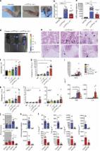
          In this study, we took advantage of human-induced pluripotent stem cells (hiPSC) and CRISPR/Cas9 technology to investigate the potential roles of RIPK1 in regulating hematopoiesis and macrophage differentiation, proinflammatory activation, and cell death pathways. Knock-out of RIPK1 in hiPSCs demonstrated that this protein is not required for erythro-myeloid differentiation. Using a well-established macrophage differentiation protocol, knock-out of RIPK1 did not block the differentiation of iPSC-derived macrophages, which displayed a similar phenotype to WT hiPSC-derived macrophages. However, knock-out of RIPK1 leads to a TNFα-dependent apoptotic death of differentiated hiPSC-derived macrophages (iPS-MΦ) and progressive loss of iPS-MΦ production irrespective of external pro-inflammatory stimuli. Live video analysis demonstrated that TLR3/4 activation of RIPK1 KO hiPSC-derived macrophages triggered TRIF and RIPK3-dependent necroptosis irrespective of caspase-8 activation. In contrast, TLR3/4 activation of WT macrophages-induced necroptosis only when caspases were inhibited, confirming the modulating effect of RIPK1 on RIPK3-mediated necroptosis through the FADD, Caspase-8 pathway. Activation of these inflammatory pathways required RIPK3 kinase activity while RIPK1 was dispensable. However, loss of RIPK1 sensitizes macrophages to activate RIPK3 in response to inflammatory stimuli, thereby exacerbating a potentially pathological inflammatory response. Taken together, these results reveal that RIPK1 has an important role in regulating the potent inflammatory pathways in authentic human macrophages that are poised to respond to external stimuli. Consequently, RIPK1 activity might be a valid target in the development of novel therapies for chronic inflammatory diseases.

          Related collections

          Most cited references24

          • Record: found
          • Abstract: found
          • Article: not found

          HIV reproducibly establishes a latent infection after acute infection of T cells in vitro.

          The presence of latent reservoirs has prevented the eradication of human immunodeficiency virus (HIV) from infected patients successfully treated with anti-retroviral therapy. The mechanism of postintegration latency is poorly understood, partly because of the lack of an in vitro model. We have used an HIV retroviral vector or a full-length HIV genome expressing green fluorescent protein to infect a T lymphocyte cell line in vitro and highly enrich for latently infected cells. HIV latency occurred reproducibly, albeit with low frequency, during an acute infection. Clonal cell lines derived from latent populations showed no detectable basal expression, but could be transcriptionally activated after treatment with phorbol esters or tumor necrosis factor alpha. Direct sequencing of integration sites demonstrated that latent clones frequently contain HIV integrated in or close to alphoid repeat elements in heterochromatin. This is in contrast to a productive infection where integration in or near heterochromatin is disfavored. These observations demonstrate that HIV can reproducibly establish a latent infection as a consequence of integration in or near heterochromatin.
            Bookmark
            • Record: found
            • Abstract: found
            • Article: found
            Is Open Access

            RIPK3 promotes cell death and NLRP3 inflammasome activation in the absence of MLKL

            RIPK3 and its substrate MLKL are essential for necroptosis, a lytic cell death proposed to cause inflammation via the release of intracellular molecules. Whether and how RIPK3 might drive inflammation in a manner independent of MLKL and cell lysis remains unclear. Here we show that following LPS treatment, or LPS-induced necroptosis, the TLR adaptor protein TRIF and inhibitor of apoptosis proteins (IAPs: X-linked IAP, cellular IAP1 and IAP2) regulate RIPK3 and MLKL ubiquitylation. Hence, when IAPs are absent, LPS triggers RIPK3 to activate caspase-8, promoting apoptosis and NLRP3–caspase-1 activation, independent of RIPK3 kinase activity and MLKL. In contrast, in the absence of both IAPs and caspase-8, RIPK3 kinase activity and MLKL are essential for TLR-induced NLRP3 activation. Consistent with in vitro experiments, interleukin-1 (IL-1)-dependent autoantibody-mediated arthritis is exacerbated in mice lacking IAPs, and is reduced by deletion of RIPK3, but not MLKL. Therefore RIPK3 can promote NLRP3 inflammasome and IL-1β inflammatory responses independent of MLKL and necroptotic cell death.
              Bookmark
              • Record: found
              • Abstract: found
              • Article: found
              Is Open Access

              Bone marrow CD169+ macrophages promote the retention of hematopoietic stem and progenitor cells in the mesenchymal stem cell niche

              Hematopoietic stem cells (HSCs) reside in specialized bone marrow (BM) niches regulated by the sympathetic nervous system (SNS). Here, we have examined whether mononuclear phagocytes modulate the HSC niche. We defined three populations of BM mononuclear phagocytes that include Gr-1hi monocytes (MOs), Gr-1lo MOs, and macrophages (MΦ) based on differential expression of Gr-1, CD115, F4/80, and CD169. Using MO and MΦ conditional depletion models, we found that reductions in BM mononuclear phagocytes led to reduced BM CXCL12 levels, the selective down-regulation of HSC retention genes in Nestin+ niche cells, and egress of HSCs/progenitors to the bloodstream. Furthermore, specific depletion of CD169+ MΦ, which spares BM MOs, was sufficient to induce HSC/progenitor egress. MΦ depletion also enhanced mobilization induced by a CXCR4 antagonist or granulocyte colony-stimulating factor. These results highlight two antagonistic, tightly balanced pathways that regulate maintenance of HSCs/progenitors in the niche during homeostasis, in which MΦ cross talk with the Nestin+ niche cell promotes retention, and in contrast, SNS signals enhance egress. Thus, strategies that target BM MΦ hold the potential to augment stem cell yields in patients that mobilize HSCs/progenitors poorly.
                Bookmark

                Author and article information

                Contributors
                william.james@path.ox.ac.uk
                jlvenero@us.es
                Journal
                Cell Death Dis
                Cell Death Dis
                Cell Death & Disease
                Nature Publishing Group UK (London )
                2041-4889
                24 September 2018
                24 September 2018
                October 2018
                : 9
                : 10
                : 973
                Affiliations
                [1 ]ISNI 0000 0004 1936 8948, GRID grid.4991.5, Sir William Dunn School of Pathology, , University of Oxford, ; Oxford, OX1 3RE UK
                [2 ]ISNI 0000 0004 1773 7922, GRID grid.414816.e, Instituto de Biomedicina de Sevilla-Hospital Universitario Virgen del Rocío/CSIC/Universidad de Sevilla, ; Sevilla, 41012 Spain
                [3 ]ISNI 0000 0001 2168 1229, GRID grid.9224.d, Departamento de Bioquímica y Biología Molecular, Facultad de Farmacia, , Universidad de Sevilla, ; Sevilla, Spain
                [4 ]ISNI 0000 0001 2353 6535, GRID grid.428999.7, Present Address: Institut Pasteur, ; 28 rue du Dr Roux, 75015 Paris, France
                Author information
                http://orcid.org/0000-0003-4790-7577
                http://orcid.org/0000-0003-2630-0442
                http://orcid.org/0000-0002-2506-1198
                http://orcid.org/0000-0003-1137-8706
                Article
                1053
                10.1038/s41419-018-1053-4
                6155173
                30250197
                fa1b35ea-db4f-4287-808c-17c193671aab
                © The Author(s) 2018

                Open Access This article is licensed under a Creative Commons Attribution 4.0 International License, which permits use, sharing, adaptation, distribution and reproduction in any medium or format, as long as you give appropriate credit to the original author(s) and the source, provide a link to the Creative Commons license, and indicate if changes were made. The images or other third party material in this article are included in the article’s Creative Commons license, unless indicated otherwise in a credit line to the material. If material is not included in the article’s Creative Commons license and your intended use is not permitted by statutory regulation or exceeds the permitted use, you will need to obtain permission directly from the copyright holder. To view a copy of this license, visit http://creativecommons.org/licenses/by/4.0/.

                History
                : 28 March 2018
                : 7 September 2018
                : 11 September 2018
                Funding
                Funded by: FundRef https://doi.org/10.13039/501100003329, Ministerio de Economía y Competitividad (Ministry of Economy and Competitiveness);
                Award ID: SAF2015-64171R
                Award Recipient :
                Funded by: FundRef https://doi.org/10.13039/100004440, Wellcome Trust;
                Award ID: WTISSF121302
                Award Recipient :
                Funded by: FundRef https://doi.org/10.13039/501100004211, Oxford Martin School, University of Oxford;
                Award ID: LC0910-004
                Award Recipient :
                Categories
                Article
                Custom metadata
                © The Author(s) 2018

                Cell biology
                Cell biology

                Comments

                Comment on this article