133
views
0
recommends
+1 Recommend
0 collections
    0
    shares
      • Record: found
      • Abstract: found
      • Article: found
      Is Open Access

      Telomerase Is Essential for Zebrafish Heart Regeneration

      research-article

      Read this article at

      Bookmark
          There is no author summary for this article yet. Authors can add summaries to their articles on ScienceOpen to make them more accessible to a non-specialist audience.

          SUMMARY

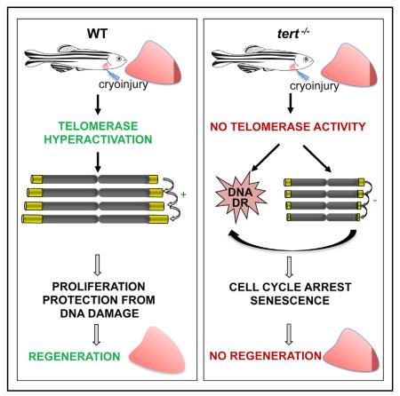
          After myocardial infarction in humans, lost cardiomyocytes are replaced by an irreversible fibrotic scar. In contrast, zebrafish hearts efficiently regenerate after injury. Complete regeneration of the zebrafish heart is driven by the strong proliferation response of its cardiomyocytes to injury. Here we show that, after cardiac injury in zebrafish, telomerase becomes hyperactivated, and telomeres elongate transiently, preceding a peak of cardiomyocyte proliferation and full organ recovery. Using a telomerase-mutant zebrafish model, we found that telomerase loss drastically decreases cardiomyocyte proliferation and fibrotic tissue regression after cryoinjury and that cardiac function does not recover. The impaired cardiomyocyte proliferation response is accompanied by the absence of cardiomyocytes with long telomeres and an increased proportion of cardiomyocytes showing DNA damage and senescence characteristics. These findings demonstrate the importance of telomerase function in heart regeneration and highlight the potential of telomerase therapy as a means of stimulating cell proliferation upon myocardial infarction.

          Graphical Abstract

          In Brief

          Bednarek et al. find that telomerase, well known for its role in elongating telomere ends, is essential during zebrafish heart regeneration. Cardiac injury hyperactivates telomerase and increases telomere length in cardiac cells. In telomerase-null mutants, cardiac cells accumulate DNA damage and do not efficiently proliferate in response to injury.

          Related collections

          Most cited references37

          • Record: found
          • Abstract: not found
          • Article: not found

          Controlling the False Discovery Rate: A Practical and Powerful Approach to Multiple Testing

            Bookmark
            • Record: found
            • Abstract: found
            • Article: not found

            The inflammatory response in myocardial injury, repair, and remodelling.

            Myocardial infarction triggers an intense inflammatory response that is essential for cardiac repair, but which is also implicated in the pathogenesis of postinfarction remodelling and heart failure. Signals in the infarcted myocardium activate toll-like receptor signalling, while complement activation and generation of reactive oxygen species induce cytokine and chemokine upregulation. Leukocytes recruited to the infarcted area, remove dead cells and matrix debris by phagocytosis, while preparing the area for scar formation. Timely repression of the inflammatory response is critical for effective healing, and is followed by activation of myofibroblasts that secrete matrix proteins in the infarcted area. Members of the transforming growth factor β family are critically involved in suppression of inflammation and activation of a profibrotic programme. Translation of these concepts to the clinic requires an understanding of the pathophysiological complexity and heterogeneity of postinfarction remodelling in patients with myocardial infarction. Individuals with an overactive and prolonged postinfarction inflammatory response might exhibit left ventricular dilatation and systolic dysfunction and might benefit from targeted anti-IL-1 or anti-chemokine therapies, whereas patients with an exaggerated fibrogenic reaction can develop heart failure with preserved ejection fraction and might require inhibition of the Smad3 (mothers against decapentaplegic homolog 3) cascade. Biomarker-based approaches are needed to identify patients with distinct pathophysiologic responses and to rationally implement inflammation-modulating strategies.
              Bookmark
              • Record: found
              • Abstract: found
              • Article: not found

              Identification of a specific telomere terminal transferase activity in Tetrahymena extracts.

              We have found a novel activity in Tetrahymena cell free extracts that adds tandem TTGGGG repeats onto synthetic telomere primers. The single-stranded DNA oligonucleotides (TTGGGG)4 and TGTGTGGGTGTGTGGGTGTGTGGG, consisting of the Tetrahymena and yeast telomeric sequences respectively, each functioned as primers for elongation, while (CCCCAA)4 and two nontelomeric sequence DNA oligomers did not. Efficient synthesis of the TTGGGG repeats depended only on addition of micromolar concentrations of oligomer primer, dGTP, and dTTP to the extract. The activity was sensitive to heat and proteinase K treatment. The repeat addition was independent of both endogenous Tetrahymena DNA and the endogenous alpha-type DNA polymerase; and a greater elongation activity was present during macronuclear development, when a large number of telomeres are formed and replicated, than during vegetative cell growth. We propose that the novel telomere terminal transferase is involved in the addition of telomeric repeats necessary for the replication of chromosome ends in eukaryotes.
                Bookmark

                Author and article information

                Journal
                101573691
                39703
                Cell Rep
                Cell Rep
                Cell reports
                2211-1247
                24 September 2015
                28 August 2015
                8 September 2015
                30 September 2015
                : 12
                : 10
                : 1691-1703
                Affiliations
                [1 ]Regeneration and Aging Group, Centro Nacional de Investigaciones Cardiovasculares (CNIC-ISCIII), Melchor Fernández Almagro 3, 28029 Madrid, Spain
                [2 ]Development of the Epicardium and Its Role during Regeneration Group, Centro Nacional de Investigaciones Cardiovasculares (CNIC-ISCIII), Melchor Fernández Almagro 3, 28029 Madrid, Spain
                [3 ]Cardiovascular Imaging in Humans, Centro Nacional de Investigaciones Cardiovasculares (CNIC-ISCIII), Melchor Fernández Almagro 3, 28029 Madrid, Spain
                [4 ]Bioinformatic Unit, Centro Nacional de Investigaciones Cardiovasculares (CNIC-ISCIII), Melchor Fernández Almagro 3, 28029 Madrid, Spain
                [5 ]Electron Microscopy Center, Complutense University, Madrid 28040, Spain
                Author notes
                [* ]Correspondence: nmercader@ 123456cnic.es (N.M.), iflores@ 123456cnic.es (I.F.)
                [6]

                Present address: Cardiovascular Research Center, Massachusetts General Hospital, Charlestown, MA 02129, USA

                [7]

                Present address: Harvard Medical School, Boston, MA 02115, USA

                [8]

                Present Address: Institute of Anatomy, University of Bern, Bern, Switzerland

                AUTHOR CONTRIBUTIONS

                D.B. performed laboratory-based experimental work, image acquisition, data quantification, data analysis, and telomere analysis. J.M.G.R. contributed to the experimental work. G.G. and J.J.B. performed and analyzed the echocardiography study. O.G.G. and T.A. and C.S.F performed the TRAP assay. I.J.M. performed the immunofluorescence analysis and cryoinjuries. M.G. contributed to the experiments with ex vivo cultures. I.D. performed experimental work. M.J.G. R. performed the transcriptome analysis. A.C. and A.Z. performed the TEM study. I.F., N.M., D.B., and J.M.G.R. designed the experiments and wrote the manuscript. I.F. directed the research.

                Article
                EMS65229
                10.1016/j.celrep.2015.07.064
                4589159
                26321646
                fc3ec025-93b6-4ad9-bc09-cf47431797ec

                This is an open access article under the CC BY-NC-ND license ( http://creativecommons.org/licenses/by-nc-nd/4.0/).

                History
                Categories
                Article

                Cell biology
                Cell biology

                Comments

                Comment on this article