91
views
0
recommends
+1 Recommend
0 collections
    0
    shares
      • Record: found
      • Abstract: found
      • Article: found
      Is Open Access

      Identification of a novel senolytic agent, navitoclax, targeting the Bcl‐2 family of anti‐apoptotic factors

      research-article

      Read this article at

          There is no author summary for this article yet. Authors can add summaries to their articles on ScienceOpen to make them more accessible to a non-specialist audience.

          Summary

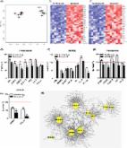
          Clearing senescent cells extends healthspan in mice. Using a hypothesis‐driven bioinformatics‐based approach, we recently identified pro‐survival pathways in human senescent cells that contribute to their resistance to apoptosis. This led to identification of dasatinib (D) and quercetin (Q) as senolytics, agents that target some of these pathways and induce apoptosis preferentially in senescent cells. Among other pro‐survival regulators identified was Bcl‐xl. Here, we tested whether the Bcl‐2 family inhibitors, navitoclax (N) and TW‐37 (T), are senolytic. Like D and Q, N is senolytic in some, but not all types of senescent cells: N reduced viability of senescent human umbilical vein epithelial cells ( HUVECs), IMR90 human lung fibroblasts, and murine embryonic fibroblasts ( MEFs), but not human primary preadipocytes, consistent with our previous finding that Bcl‐xl si RNA is senolytic in HUVECs, but not preadipocytes. In contrast, T had little senolytic activity. N targets Bcl‐2, Bcl‐xl, and Bcl‐w, while T targets Bcl‐2, Bcl‐xl, and Mcl‐1. The combination of Bcl‐2, Bcl‐xl, and Bcl‐w si RNAs was senolytic in HUVECs and IMR90 cells, while combination of Bcl‐2, Bcl‐xl, and Mcl‐1 si RNAs was not. Susceptibility to N correlated with patterns of Bcl‐2 family member proteins in different types of human senescent cells, as has been found in predicting response of cancers to N. Thus, N is senolytic and acts in a potentially predictable cell type‐restricted manner. The hypothesis‐driven, bioinformatics‐based approach we used to discover that dasatinib (D) and quercetin (Q) are senolytic can be extended to increase the repertoire of senolytic drugs, including additional cell type‐specific senolytic agents.

          Related collections

          Most cited references21

          • Record: found
          • Abstract: found
          • Article: found
          Is Open Access

          The Achilles’ heel of senescent cells: from transcriptome to senolytic drugs

          The healthspan of mice is enhanced by killing senescent cells using a transgenic suicide gene. Achieving the same using small molecules would have a tremendous impact on quality of life and the burden of age-related chronic diseases. Here, we describe the rationale for identification and validation of a new class of drugs termed senolytics, which selectively kill senescent cells. By transcript analysis, we discovered increased expression of pro-survival networks in senescent cells, consistent with their established resistance to apoptosis. Using siRNA to silence expression of key nodes of this network, including ephrins (EFNB1 or 3), PI3Kδ, p21, BCL-xL, or plasminogen-activated inhibitor-2, killed senescent cells, but not proliferating or quiescent, differentiated cells. Drugs targeting these same factors selectively killed senescent cells. Dasatinib eliminated senescent human fat cell progenitors, while quercetin was more effective against senescent human endothelial cells and mouse BM-MSCs. The combination of dasatinib and quercetin was effective in eliminating senescent MEFs. In vivo, this combination reduced senescent cell burden in chronologically aged, radiation-exposed, and progeroid Ercc1 −/Δ mice. In old mice, cardiac function and carotid vascular reactivity were improved 5 days after a single dose. Following irradiation of one limb in mice, a single dose led to improved exercise capacity for at least 7 months following drug treatment. Periodic drug administration extended healthspan in Ercc1 −/Δ mice, delaying age-related symptoms and pathology, osteoporosis, and loss of intervertebral disk proteoglycans. These results demonstrate the feasibility of selectively ablating senescent cells and the efficacy of senolytics for alleviating symptoms of frailty and extending healthspan.
            Bookmark
            • Record: found
            • Abstract: found
            • Article: not found

            Senescence and tumour clearance is triggered by p53 restoration in murine liver carcinomas.

            Although cancer arises from a combination of mutations in oncogenes and tumour suppressor genes, the extent to which tumour suppressor gene loss is required for maintaining established tumours is poorly understood. p53 is an important tumour suppressor that acts to restrict proliferation in response to DNA damage or deregulation of mitogenic oncogenes, by leading to the induction of various cell cycle checkpoints, apoptosis or cellular senescence. Consequently, p53 mutations increase cell proliferation and survival, and in some settings promote genomic instability and resistance to certain chemotherapies. To determine the consequences of reactivating the p53 pathway in tumours, we used RNA interference (RNAi) to conditionally regulate endogenous p53 expression in a mosaic mouse model of liver carcinoma. We show that even brief reactivation of endogenous p53 in p53-deficient tumours can produce complete tumour regressions. The primary response to p53 was not apoptosis, but instead involved the induction of a cellular senescence program that was associated with differentiation and the upregulation of inflammatory cytokines. This program, although producing only cell cycle arrest in vitro, also triggered an innate immune response that targeted the tumour cells in vivo, thereby contributing to tumour clearance. Our study indicates that p53 loss can be required for the maintenance of aggressive carcinomas, and illustrates how the cellular senescence program can act together with the innate immune system to potently limit tumour growth.
              Bookmark
              • Record: found
              • Abstract: found
              • Article: not found

              Control of apoptosis by p53.

              The p53 tumor suppressor acts to integrate multiple stress signals into a series of diverse antiproliferative responses. One of the most important p53 functions is its ability to activate apoptosis, and disruption of this process can promote tumor progression and chemoresistance. p53 apparently promotes apoptosis through transcription-dependent and -independent mechanisms that act in concert to ensure that the cell death program proceeds efficiently. Moreover, the apoptotic activity of p53 is tightly controlled, and is influenced by a series of quantitative and qualitative events that influence the outcome of p53 activation. Interestingly, other p53 family members can also promote apoptosis, either in parallel or in concert with p53. Although incomplete, our current understanding of p53 illustrates how apoptosis can be integrated into a larger tumor suppressor network controlled by different signals, environmental factors, and cell type. Understanding this network in more detail will provide insights into cancer and other diseases, and will identify strategies to improve their therapeutic treatment.
                Bookmark

                Author and article information

                Journal
                Aging Cell
                Aging Cell
                10.1111/(ISSN)1474-9726
                ACEL
                Aging Cell
                John Wiley and Sons Inc. (Hoboken )
                1474-9718
                1474-9726
                18 March 2016
                June 2016
                : 15
                : 3 ( doiID: 10.1111/acel.2016.15.issue-3 )
                : 428-435
                Affiliations
                [ 1 ] Robert and Arlene Kogod Center on AgingMayo Clinic Rochester MNUSA
                [ 2 ] Department of Metabolism and AgingThe Scripps Research Institute Jupiter FLUSA
                [ 3 ] Center of Medical Physics and TechnologyHefei Institutes of Physical Sciences HefeiChina
                [ 4 ] Arthritis and Clinical Immunology Research ProgramOklahoma Medical Research Foundation Oklahoma City OKUSA
                Author notes
                [*] [* ] Correspondence

                James L. Kirkland, Robert and Arlene Kogod Center on Aging, Mayo Clinic, 200 First Street, S.W., Rochester, MN 55905, USA. Tel.: +1 507 266 9151; fax: +1 507 293 3853; e‐mail: Kirkland.james@ 123456mayo.edu

                [†]

                Co‐first authors

                Article
                ACEL12445
                10.1111/acel.12445
                4854923
                26711051
                94269170-6533-4ec0-ae5f-975b9f740b2e
                © 2015 The Authors. Aging Cell published by the Anatomical Society and John Wiley & Sons Ltd.

                This is an open access article under the terms of the Creative Commons Attribution License, which permits use, distribution and reproduction in any medium, provided the original work is properly cited.

                History
                : 29 December 2015
                Page count
                Pages: 8
                Funding
                Funded by: NIH
                Award ID: AG13925
                Award ID: DK50456
                Award ID: AG043376
                Funded by: Noaber, Ellison
                Funded by: Ted Nash and Glenn Foundations
                Funded by: American Federation for Aging Research
                Funded by: Connor Group
                Funded by: Natural Science Foundation of China
                Award ID: 81572948
                Categories
                Original Article
                Original Articles
                Custom metadata
                2.0
                acel12445
                June 2016
                Converter:WILEY_ML3GV2_TO_NLMPMC version:4.9.1 mode:remove_FC converted:10.06.2016

                Cell biology
                abt‐263,bcl‐2 family,dasatinib,quercetin,senescent cells,tw‐37
                Cell biology
                abt‐263, bcl‐2 family, dasatinib, quercetin, senescent cells, tw‐37

                Comments

                Comment on this article