152
views
0
recommends
+1 Recommend
0 collections
    0
    shares
      • Record: found
      • Abstract: found
      • Article: found
      Is Open Access

      Quantitative OCT angiography of optic nerve head blood flow

      research-article

      Read this article at

      Bookmark
          There is no author summary for this article yet. Authors can add summaries to their articles on ScienceOpen to make them more accessible to a non-specialist audience.

          Abstract

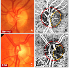
          Optic nerve head (ONH) blood flow may be associated with glaucoma development. A reliable method to quantify ONH blood flow could provide insight into the vascular component of glaucoma pathophysiology. Using ultrahigh-speed optical coherence tomography (OCT), we developed a new 3D angiography algorithm called split-spectrum amplitude-decorrelation angiography (SSADA) for imaging ONH microcirculation. In this study, a method to quantify SSADA results was developed and used to detect ONH perfusion changes in early glaucoma. En face maximum projection was used to obtain 2D disc angiograms, from which the average decorrelation values (flow index) and the percentage area occupied by vessels (vessel density) were computed from the optic disc and a selected region within it. Preperimetric glaucoma patients had significant reductions of ONH perfusion compared to normals. This pilot study indicates OCT angiography can detect the abnormalities of ONH perfusion and has the potential to reveal the ONH blood flow mechanism related to glaucoma.

          Related collections

          Most cited references34

          • Record: found
          • Abstract: found
          • Article: not found

          Optical coherence tomography.

          A technique called optical coherence tomography (OCT) has been developed for noninvasive cross-sectional imaging in biological systems. OCT uses low-coherence interferometry to produce a two-dimensional image of optical scattering from internal tissue microstructures in a way that is analogous to ultrasonic pulse-echo imaging. OCT has longitudinal and lateral spatial resolutions of a few micrometers and can detect reflected signals as small as approximately 10(-10) of the incident optical power. Tomographic imaging is demonstrated in vitro in the peripapillary area of the retina and in the coronary artery, two clinically relevant examples that are representative of transparent and turbid media, respectively.
            Bookmark
            • Record: found
            • Abstract: found
            • Article: found
            Is Open Access

            Split-spectrum amplitude-decorrelation angiography with optical coherence tomography

            Amplitude decorrelation measurement is sensitive to transverse flow and immune to phase noise in comparison to Doppler and other phase-based approaches. However, the high axial resolution of OCT makes it very sensitive to the pulsatile bulk motion noise in the axial direction. To overcome this limitation, we developed split-spectrum amplitude-decorrelation angiography (SSADA) to improve the signal-to-noise ratio (SNR) of flow detection. The full OCT spectrum was split into several narrower bands. Inter-B-scan decorrelation was computed using the spectral bands separately and then averaged. The SSADA algorithm was tested on in vivo images of the human macula and optic nerve head. It significantly improved both SNR for flow detection and connectivity of microvascular network when compared to other amplitude-decorrelation algorithms.
              Bookmark
              • Record: found
              • Abstract: found
              • Article: not found

              Speckle variance detection of microvasculature using swept-source optical coherence tomography.

              We report on imaging of microcirculation by calculating the speckle variance of optical coherence tomography (OCT) structural images acquired using a Fourier domain mode-locked swept-wavelength laser. The algorithm calculates interframe speckle variance in two-dimensional and three-dimensional OCT data sets and shows little dependence to the Doppler angle ranging from 75 degrees to 90 degrees . We demonstrate in vivo detection of blood flow in vessels as small as 25 microm in diameter in a dorsal skinfold window chamber model with direct comparison with intravital fluorescence confocal microscopy. This technique can visualize vessel-size-dependent vascular shutdown and transient vascular occlusion during Visudyne photodynamic therapy and may provide opportunities for studying therapeutic effects of antivascular treatments without on exogenous contrast agent.
                Bookmark

                Author and article information

                Journal
                Biomed Opt Express
                Biomed Opt Express
                BOE
                Biomedical Optics Express
                Optical Society of America
                2156-7085
                07 November 2012
                01 December 2012
                07 November 2012
                : 3
                : 12
                : 3127-3137
                Affiliations
                [1 ]Casey Eye Institute, Oregon Health & Science University, Portland, OR 97239, USA
                [2 ]Department of Electrical Engineering, University of Southern California, Los Angeles, CA 90089, USA
                [3 ]Department of Electrical Engineering and Computer Science, and Research Laboratory of Electronics, Massachusetts Institute of Technology, Cambridge, MA 02139, USA
                Author notes
                Article
                173662
                10.1364/BOE.3.003127
                3521313
                23243564
                196fb690-93a6-4b98-ab45-28e338f7be29
                ©2012 Optical Society of America

                This is an open-access article distributed under the terms of the Creative Commons Attribution-Noncommercial-No Derivative Works 3.0 Unported License, which permits download and redistribution, provided that the original work is properly cited. This license restricts the article from being modified or used commercially.

                History
                : 1 August 2011
                : 11 September 2012
                : 15 October 2012
                Funding
                Funded by: NIH
                Award ID: R01 EY013516
                Award ID: R01-EY11289-26
                Funded by: AFOSR
                Award ID: FA9550-10-1-0551
                Categories
                Ophthalmology Applications
                Custom metadata
                True
                0

                Vision sciences
                (170.4470) ophthalmology,(170.4500) optical coherence tomography,(170.3880) medical and biological imaging

                Comments

                Comment on this article