61
views
0
recommends
+1 Recommend
0 collections
    0
    shares
      • Record: found
      • Abstract: found
      • Article: found
      Is Open Access

      Age-Associated Sperm DNA Methylation Alterations: Possible Implications in Offspring Disease Susceptibility

      research-article

      Read this article at

      Bookmark
          There is no author summary for this article yet. Authors can add summaries to their articles on ScienceOpen to make them more accessible to a non-specialist audience.

          Abstract

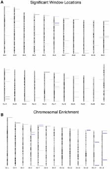
          Recent evidence demonstrates a role for paternal aging on offspring disease susceptibility. It is well established that various neuropsychiatric disorders (schizophrenia, autism, etc.), trinucleotide expansion associated diseases (myotonic dystrophy, Huntington's, etc.) and even some forms of cancer have increased incidence in the offspring of older fathers. Despite strong epidemiological evidence that these alterations are more common in offspring sired by older fathers, in most cases the mechanisms that drive these processes are unclear. However, it is commonly believed that epigenetics, and specifically DNA methylation alterations, likely play a role. In this study we have investigated the impact of aging on DNA methylation in mature human sperm. Using a methylation array approach we evaluated changes to sperm DNA methylation patterns in 17 fertile donors by comparing the sperm methylome of 2 samples collected from each individual 9–19 years apart. With this design we have identified 139 regions that are significantly and consistently hypomethylated with age and 8 regions that are significantly hypermethylated with age. A representative subset of these alterations have been confirmed in an independent cohort. A total of 117 genes are associated with these regions of methylation alterations (promoter or gene body). Intriguingly, a portion of the age-related changes in sperm DNA methylation are located at genes previously associated with schizophrenia and bipolar disorder. While our data does not establish a causative relationship, it does raise the possibility that the age-associated methylation of the candidate genes that we observe in sperm might contribute to the increased incidence of neuropsychiatric and other disorders in the offspring of older males. However, further study is required to determine whether, and to what extent, a causative relationship exists.

          Author Summary

          There is a striking trend of delayed parenthood in developed countries due to secular and socioeconomic pressures. As a result, physicians commonly consult with concerned patients inquiring about the impact of advanced age on their ability to conceive healthy offspring. The concern has more frequently surrounded the effects of advanced maternal age, but recent evidence suggests negative effects of advanced paternal age as well. Specifically, studies have demonstrated increased incidence of neuropsychiatric and other disorders in the offspring of older males. In this study we have investigated a commonly hypothesized mechanism for this effect, namely sperm DNA methylation alteration. Our data indicate that specific genomic regions of DNA methylation are commonly altered with age, suggesting that some regions of the sperm genome are more susceptible than others to age-related epigenetic changes. Importantly, a significant portion of these alterations occur at genes known to be associated with schizophrenia and bipolar disorder, both of which display increased incidence in the offspring of older fathers. These data will be important in driving future studies aimed at determining the impact that these methylation alterations may have on offspring health and will thus enable couples at advanced reproductive ages to be more informed of possible risks.

          Related collections

          Most cited references38

          • Record: found
          • Abstract: found
          • Article: not found

          DNA methylation age of human tissues and cell types

          Background It is not yet known whether DNA methylation levels can be used to accurately predict age across a broad spectrum of human tissues and cell types, nor whether the resulting age prediction is a biologically meaningful measure. Results I developed a multi-tissue predictor of age that allows one to estimate the DNA methylation age of most tissues and cell types. The predictor, which is freely available, was developed using 8,000 samples from 82 Illumina DNA methylation array datasets, encompassing 51 healthy tissues and cell types. I found that DNA methylation age has the following properties: first, it is close to zero for embryonic and induced pluripotent stem cells; second, it correlates with cell passage number; third, it gives rise to a highly heritable measure of age acceleration; and, fourth, it is applicable to chimpanzee tissues. Analysis of 6,000 cancer samples from 32 datasets showed that all of the considered 20 cancer types exhibit significant age acceleration, with an average of 36 years. Low age-acceleration of cancer tissue is associated with a high number of somatic mutations and TP53 mutations, while mutations in steroid receptors greatly accelerate DNA methylation age in breast cancer. Finally, I characterize the 353 CpG sites that together form an aging clock in terms of chromatin states and tissue variance. Conclusions I propose that DNA methylation age measures the cumulative effect of an epigenetic maintenance system. This novel epigenetic clock can be used to address a host of questions in developmental biology, cancer and aging research.
            Bookmark
            • Record: found
            • Abstract: found
            • Article: not found

            The relationship between trinucleotide (CAG) repeat length and clinical features of Huntington's disease.

            Huntington's disease (HD) is associated with the expansion of a CAG trinucleotide repeat in a novel gene. We have assessed 360 HD individuals from 259 unrelated families and found a highly significant correlation (r = 0.70, p = 10(-7)) between the age of onset and the repeat length, which accounts for approximately 50% of the variation in the age of onset. Significant associations were also found between repeat length and age of death and onset of other clinical features. Sib pair and parent-child analysis revealed that the CAG repeat demonstrates only mild instability. Affected HD siblings had significant correlations for trinucleotide expansion (r = 0.66, p < 0.001) which was not apparent for affected parent-child pairs.
              Bookmark
              • Record: found
              • Abstract: found
              • Article: not found

              Telomere length predicts replicative capacity of human fibroblasts.

              When human fibroblasts from different donors are grown in vitro, only a small fraction of the variation in their finite replicative capacity is explained by the chronological age of the donor. Because we had previously shown that telomeres, the terminal guanine-rich sequences of chromosomes, shorten throughout the life-span of cultured cells, we wished to determine whether variation in initial telomere length would account for the unexplained variation in replicative capacity. Analysis of cells from 31 donors (aged 0-93 yr) indicated relatively weak correlations between proliferative ability and donor age (m = -0.2 doubling per yr; r = -0.42; P = 0.02) and between telomeric DNA and donor age (m = -15 base pairs per yr; r = -0.43; P = 0.02). However, there was a striking correlation, valid over the entire age range of the donors, between replicative capacity and initial telomere length (m = 10 doublings per kilobase pair; r = 0.76; P = 0.004), indicating that cell strains with shorter telomeres underwent significantly fewer doublings than those with longer telomeres. These observations suggest that telomere length is a biomarker of somatic cell aging in humans and are consistent with a causal role for telomere loss in this process. We also found that fibroblasts from Hutchinson-Gilford progeria donors had short telomeres, consistent with their reduced division potential in vitro. In contrast, telomeres from sperm DNA did not decrease with age of the donor, suggesting that a mechanism for maintaining telomere length, such as telomerase expression, may be active in germ-line tissue.
                Bookmark

                Author and article information

                Contributors
                Role: Editor
                Journal
                PLoS Genet
                PLoS Genet
                plos
                plosgen
                PLoS Genetics
                Public Library of Science (San Francisco, USA )
                1553-7390
                1553-7404
                July 2014
                10 July 2014
                : 10
                : 7
                : e1004458
                Affiliations
                [1 ]Andrology and IVF Laboratories, Department of Surgery, University of Utah School of Medicine, Salt Lake City, Utah, United States of America
                [2 ]Department of Oncological Sciences, Huntsman Cancer Institute, University of Utah School of Medicine, Salt Lake City, Utah, United States of America
                [3 ]Howard Hughes Medical Institute, Chevy Chase, Maryland, United States of America
                [4 ]Department of Genetics, University of Utah School of Medicine, Salt Lake City, Utah, United States of America
                [5 ]Department of Obstetrics and Gynecology, University of Utah School of Medicine, Salt Lake City, Utah, United States of America
                Albert Einstein College of Medicine, United States of America
                Author notes

                The authors have declared that no competing interests exist.

                Conceived and designed the experiments: DTC BRC TGJ KIA. Performed the experiments: TGJ KIA. Analyzed the data: CP TGJ BRC DTC KIA. Contributed reagents/materials/analysis tools: DTC BRC. Wrote the paper: TGJ CP BRC DTC KIA.

                Article
                PGENETICS-D-13-03189
                10.1371/journal.pgen.1004458
                4091790
                25010591
                1fd45a5a-94da-4edf-931d-c5a9411c3d67
                Copyright @ 2014

                This is an open-access article distributed under the terms of the Creative Commons Attribution License, which permits unrestricted use, distribution, and reproduction in any medium, provided the original author and source are credited.

                History
                : 21 November 2013
                : 9 May 2014
                Page count
                Pages: 13
                Funding
                An internal University of Utah small grant from the “University of Utah Center on Aging” was used for this study. Additionally, clinical funds were used for this study. No outside grant agency funds were applied. The funders had no role in study design, data collection and analysis, decision to publish, or preparation of the manuscript.
                Categories
                Research Article
                Biology and Life Sciences
                Genetics
                Epigenetics
                Medicine and Health Sciences
                Urology
                Infertility

                Genetics
                Genetics

                Comments

                Comment on this article