52
views
0
recommends
+1 Recommend
0 collections
    0
    shares
      • Record: found
      • Abstract: found
      • Article: found
      Is Open Access

      Conformational plasticity of RNA for target recognition as revealed by the 2.15 Å crystal structure of a human IgG–aptamer complex

      research-article

      Read this article at

      Bookmark
          There is no author summary for this article yet. Authors can add summaries to their articles on ScienceOpen to make them more accessible to a non-specialist audience.

          Abstract

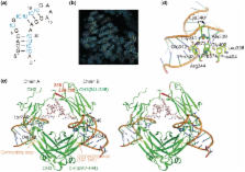
          Aptamers are short single-stranded nucleic acids with high affinity to target molecules and are applicable to therapeutics and diagnostics. Regardless of an increasing number of reported aptamers, the structural basis of the interaction of RNA aptamer with proteins is poorly understood. Here, we determined the 2.15 Å crystal structure of the Fc fragment of human IgG1 (hFc1) complexed with an anti-Fc RNA aptamer. The aptamer adopts a characteristic structure fit to hFc1 that is stabilized by a calcium ion, and the binding activity of the aptamer can be controlled many times by calcium chelation and addition. Importantly, the aptamer–hFc1 interaction involves mainly van der Waals contacts and hydrogen bonds rather than electrostatic forces, in contrast to other known aptamer–protein complexes. Moreover, the aptamer–hFc1 interaction involves human IgG-specific amino acids, rendering the aptamer specific to human IgGs, and not crossreactive to other species IgGs. Hence, the aptamer is a potent alternative for protein A affinity purification of Fc-fusion proteins and therapeutic antibodies. These results demonstrate, from a structural viewpoint, that conformational plasticity and selectivity of an RNA aptamer is achieved by multiple interactions other than electrostatic forces, which is applicable to many protein targets of low or no affinity to nucleic acids.

          Related collections

          Most cited references39

          • Record: found
          • Abstract: found
          • Article: not found

          Systematic evolution of ligands by exponential enrichment: RNA ligands to bacteriophage T4 DNA polymerase.

          L Gold, C Tuerk (1990)
          High-affinity nucleic acid ligands for a protein were isolated by a procedure that depends on alternate cycles of ligand selection from pools of variant sequences and amplification of the bound species. Multiple rounds exponentially enrich the population for the highest affinity species that can be clonally isolated and characterized. In particular one eight-base region of an RNA that interacts with the T4 DNA polymerase was chosen and randomized. Two different sequences were selected by this procedure from the calculated pool of 65,536 species. One is the wild-type sequence found in the bacteriophage mRNA; one is varied from wild type at four positions. The binding constants of these two RNA's to T4 DNA polymerase are equivalent. These protocols with minimal modification can yield high-affinity ligands for any protein that binds nucleic acids as part of its function; high-affinity ligands could conceivably be developed for any target molecule.
            Bookmark
            • Record: found
            • Abstract: found
            • Article: not found

            Structure of the human telomerase RNA pseudoknot reveals conserved tertiary interactions essential for function.

            Human telomerase contains a 451 nt RNA (hTR) and several proteins, including a specialized reverse transcriptase (hTERT). The 5' half of hTR comprises the pseudoknot (core) domain, which includes the RNA template for telomere synthesis and a highly conserved pseudoknot that is required for telomerase activity. The solution structure of this essential pseudoknot, presented here, reveals an extended triple helix surrounding the helical junction. The network of tertiary interactions explains the phylogenetic sequence conservation and existing human and mouse TR functional studies as well as mutations linked to disease. Thermodynamic stability, dimerization potential, and telomerase activity of mutant RNAs that alter the tertiary contacts were investigated. Telomerase activity is strongly correlated with tertiary structure stability, whereas there is no correlation with dimerization potential of the pseudoknot. These studies reveal that a conserved pseudoknot tertiary structure is required for telomerase activity.
              Bookmark
              • Record: found
              • Abstract: found
              • Article: not found

              Three-dimensional structure of a hammerhead ribozyme.

              The hammerhead ribozyme is a small catalytic RNA motif made up of three base-paired stems and a core of highly conserved, non-complementary nucleotides essential for catalysis. The X-ray crystallographic structure of a hammerhead RNA-DNA ribozyme-inhibitor complex at 2.6 A resolution reveals that the base-paired stems are A-form helices and that the core has two structural domains. The first domain is formed by the sequence 5'-CUGA following stem I and is a sharp turn identical to the uridine turn of transfer RNA, whereas the second is a non-Watson-Crick three-base-pair duplex with a divalent-ion binding site. The phosphodiester backbone of the DNA inhibitor strand is splayed out at the phosphate 5' to the cleavage site. The structure indicates that the ribozyme may destabilize a substrate strand in order to facilitate twisting of the substrate to allow cleavage of the scissile bond.
                Bookmark

                Author and article information

                Journal
                Nucleic Acids Res
                nar
                nar
                Nucleic Acids Research
                Oxford University Press
                0305-1048
                1362-4962
                November 2010
                November 2010
                30 July 2010
                30 July 2010
                : 38
                : 21
                : 7822-7829
                Affiliations
                1Department of Life and Environmental Sciences, Faculty of Engineering, Chiba Institute of Technology, Narashino-shi, Chiba 275-0016, 2CREST JST, Minato-ku, Tokyo 108-8639, 3Graduate School of Engineering, Osaka University, 4CREST JST, Suita, Osaka 565-0871, 5Ribomic Inc., 3-16-13 Shirokanedai, Minato-ku, Tokyo 108-0071, 6SOSHO Inc., Osaka 541-0053, 7Graduate School of Bioscience and Biotechnology, Tokyo Institute of Technology, Nagatsuta, Midori-ku, Yokohama 226-8501 and 8Department of Basic Medical Sciences, Institute of Medical Science, University of Tokyo, Minato-ku, Tokyo 108-8639, Japan
                Author notes
                *To whom correspondence should be addressed. Tel: +81 6 6879 7410; Fax: +81 6 6879 7409; Email: matsumura@ 123456chem.eng.osaka-u.ac.jp
                Correspondence may also be addressed to Yoshikazu Nakamura. Tel: +81 3 5449 5307; Fax: +81 3 5449 5415; Email: nak@ 123456ims.u-tokyo.ac.jp

                The authors wish it to be known that, in their opinion, the first two authors should be regarded as joint First Authors.

                Article
                gkq615
                10.1093/nar/gkq615
                2995045
                20675355
                509b65ea-71aa-4516-b881-f59e7cdb20e3
                © The Author(s) 2010. Published by Oxford University Press.

                This is an Open Access article distributed under the terms of the Creative Commons Attribution Non-Commercial License ( http://creativecommons.org/licenses/by-nc/2.5), which permits unrestricted non-commercial use, distribution, and reproduction in any medium, provided the original work is properly cited.

                History
                : 17 May 2010
                : 20 June 2010
                : 24 June 2010
                Categories
                Structural Biology

                Genetics
                Genetics

                Comments

                Comment on this article