14
views
0
recommends
+1 Recommend
0 collections
    0
    shares
      • Record: found
      • Abstract: found
      • Article: found
      Is Open Access

      BCL-2 family isoforms in apoptosis and cancer

      review-article

      Read this article at

      Bookmark
          There is no author summary for this article yet. Authors can add summaries to their articles on ScienceOpen to make them more accessible to a non-specialist audience.

          Abstract

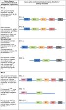
          The BCl-2 family has long been identified for its role in apoptosis. Following the initial discovery of BCL-2 in the context of B-cell lymphoma in the 1980s, a number of homologous proteins have since been identified. The members of the Bcl-2 family are designated as such due to their BCL-2 homology (BH) domains and involvement in apoptosis regulation. The BH domains facilitate the family members’ interactions with each other and can indicate pro- or anti-apoptotic function. Traditionally, these proteins are categorised into one of the three subfamilies; anti-apoptotic, BH3-only (pro-apoptotic), and pore-forming or ‘executioner’ (pro-apoptotic) proteins. Each of the BH3-only or anti-apoptotic proteins has a distinct pattern of activation, localisation and response to cell death or survival stimuli. All of these can vary across cell or stress types, or developmental stage, and this can cause the delineation of the roles of BCL-2 family members. Added to this complexity is the presence of relatively uncharacterised isoforms of many of the BCL-2 family members. There is a gap in our knowledge regarding the function of BCL-2 family isoforms. BH domain status is not always predictive or indicative of protein function, and several other important sequences, which can contribute to apoptotic activity have been identified. While therapeutic strategies targeting the BCL-2 family are constantly under development, it is imperative that we understand the molecules, which we are attempting to target. This review, discusses our current knowledge of anti-apoptotic BCL-2 family isoforms. With significant improvements in the potential for splicing therapies, it is important that we begin to understand the distinctions of the BCL-2 family, not limited to just the mechanisms of apoptosis control, but in their roles outside of apoptosis.

          Related collections

          Most cited references131

          • Record: found
          • Abstract: found
          • Article: not found

          Bcl-2 heterodimerizes in vivo with a conserved homolog, Bax, that accelerates programmed cell death.

          Bcl-2 protein is able to repress a number of apoptotic death programs. To investigate the mechanism of Bcl-2's effect, we examined whether Bcl-2 interacted with other proteins. We identified an associated 21 kd protein partner, Bax, that has extensive amino acid homology with Bcl-2, focused within highly conserved domains I and II. Bax is encoded by six exons and demonstrates a complex pattern of alternative RNA splicing that predicts a 21 kd membrane (alpha) and two forms of cytosolic protein (beta and gamma). Bax homodimerizes and forms heterodimers with Bcl-2 in vivo. Overexpressed Bax accelerates apoptotic death induced by cytokine deprivation in an IL-3-dependent cell line. Overexpressed Bax also counters the death repressor activity of Bcl-2. These data suggest a model in which the ratio of Bcl-2 to Bax determines survival or death following an apoptotic stimulus.
            Bookmark
            • Record: found
            • Abstract: found
            • Article: not found

            Direct activation of Bax by p53 mediates mitochondrial membrane permeabilization and apoptosis.

            The tumor suppressor p53 exerts its anti-neoplastic activity primarily through the induction of apoptosis. We found that cytosolic localization of endogenous wild-type or trans-activation-deficient p53 was necessary and sufficient for apoptosis. p53 directly activated the proapoptotic Bcl-2 protein Bax in the absence of other proteins to permeabilize mitochondria and engage the apoptotic program. p53 also released both proapoptotic multidomain proteins and BH3-only proteins [Proapoptotic Bcl-2 family proteins that share only the third Bcl-2 homology domain (BH3)] that were sequestered by Bcl-xL. The transcription-independent activation of Bax by p53 occurred with similar kinetics and concentrations to those produced by activated Bid. We propose that when p53 accumulates in the cytosol, it can function analogously to the BH3-only subset of proapoptotic Bcl-2 proteins to activate Bax and trigger apoptosis.
              Bookmark
              • Record: found
              • Abstract: found
              • Article: not found

              Bcl-2 gene promotes haemopoietic cell survival and cooperates with c-myc to immortalize pre-B cells.

              A common feature of follicular lymphoma, the most prevalent haematological malignancy in humans, is a chromosome translocation (t(14;18] that has coupled the immunoglobulin heavy chain locus to a chromosome 18 gene denoted bcl-2. By analogy with the translocated c-myc oncogene in other B-lymphoid tumours bcl-2 is a candidate oncogene, but no biological effects of bcl-2 have yet been reported. To test whether bcl-2 influences the growth of haematopoietic cells, either alone or together with a deregulated c-myc gene, we have introduced a human bcl-2 complementary DNA using a retroviral vector into bone marrow cells from either normal or E mu-myc transgenic mice, in which B-lineage cells constitutively express the c-myc gene. Bcl-2 cooperated with c-myc to promote proliferation of B-cell precursors, some of which became tumorigenic. To determine how bcl-2 expression impinges on growth factor requirements, the gene was introduced into a lymphoid and a myeloid cell line that require interleukin 3 (IL-3). In the absence of IL-3, bcl-2 promoted the survival of the infected cells but they persisted in a G0 state, rather than proliferating. These results argue that bcl-2 provided a distinct survival signal to the cell and may contribute to neoplasia by allowing a clone to persist until other oncogenes, such as c-myc, become activated.
                Bookmark

                Author and article information

                Contributors
                Nikola.bowden@newcastle.edu.au
                Journal
                Cell Death Dis
                Cell Death Dis
                Cell Death & Disease
                Nature Publishing Group UK (London )
                2041-4889
                21 February 2019
                21 February 2019
                March 2019
                : 10
                : 3
                : 177
                Affiliations
                [1 ]ISNI 0000 0000 8831 109X, GRID grid.266842.c, Faculty of Health and Medicine, School of Biomedical Sciences and Pharmacy, , University of Newcastle, ; Newcastle, NSW Australia
                [2 ]GRID grid.413648.c, Hunter Medical Research Institute, ; Newcastle, NSW Australia
                [3 ]ISNI 0000 0000 8831 109X, GRID grid.266842.c, Faculty of Health and Medicine, School of Medicine and Public Health, , University of Newcastle, ; Newcastle, NSW Australia
                Author information
                http://orcid.org/0000-0002-6047-1694
                Article
                1407
                10.1038/s41419-019-1407-6
                6384907
                30792387
                5adb8958-7aa9-4cc4-a26a-751cd7cb12b6
                © The Author(s) 2019

                Open Access This article is licensed under a Creative Commons Attribution 4.0 International License, which permits use, sharing, adaptation, distribution and reproduction in any medium or format, as long as you give appropriate credit to the original author(s) and the source, provide a link to the Creative Commons license, and indicate if changes were made. The images or other third party material in this article are included in the article’s Creative Commons license, unless indicated otherwise in a credit line to the material. If material is not included in the article’s Creative Commons license and your intended use is not permitted by statutory regulation or exceeds the permitted use, you will need to obtain permission directly from the copyright holder. To view a copy of this license, visit http://creativecommons.org/licenses/by/4.0/.

                History
                : 23 September 2018
                : 17 December 2018
                : 29 January 2019
                Funding
                Funded by: FundRef https://doi.org/10.13039/501100001171, Cancer Institute NSW (Cancer Institute New South Wales);
                Award ID: 14CDF117
                Award Recipient :
                Funded by: FundRef https://doi.org/10.13039/501100001081, Hunter Medical Research Institute (HMRI);
                Funded by: University of Newcastle Priority Research Centre for Cancer Research Innovation and Translation
                Categories
                Review Article
                Custom metadata
                © The Author(s) 2019

                Cell biology
                Cell biology

                Comments

                Comment on this article