14
views
0
recommends
+1 Recommend
0 collections
    0
    shares
      • Record: found
      • Abstract: found
      • Article: not found

      Ribosomal genes in focus : new transcripts label the dense fibrillar components and form clusters indicative of “Christmas trees” in situ

      research-article

      Read this article at

      ScienceOpenPublisherPMC
      Bookmark
          There is no author summary for this article yet. Authors can add summaries to their articles on ScienceOpen to make them more accessible to a non-specialist audience.

          Abstract

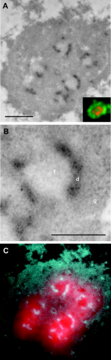
          T he organization of transcriptionally active ribosomal genes in animal cell nucleoli is investigated in this study in order to address the long-standing controversy with regard to the intranucleolar localization of these genes. Detailed analyses of HeLa cell nucleoli include direct localization of ribosomal genes by in situ hybridization and their indirect localization via nascent ribosomal transcript mappings. On the light microscopy (LM) level, ribosomal genes map in 10–40 fluorescence foci per nucleus, and transcription activity is associated with most foci. We demonstrate that each nucleolar focus observed by LM corresponds, on the EM level, to an individual fibrillar center (FC) and surrounding dense fibrillar components (DFCs). The EM data identify the DFC as the nucleolar subcompartment in which rRNA synthesis takes place, consistent with detection of rDNA within the DFC. The highly sensitive method for mapping nascent transcripts in permeabilized cells on ultrastructural level provides intense and unambiguous clustered immunogold signal over the DFC, whereas very little to no label is detected over the FC. This signal is strongly indicative of nascent “Christmas trees” of rRNA associated with individual rDNA genes, sampled on the surface of thin sections. Stereological analysis of the clustered transcription signal further suggests that these Christmas trees may be contorted in space and exhibit a DNA compaction ratio on the order of 4–5.5.

          Related collections

          Most cited references17

          • Record: found
          • Abstract: found
          • Article: not found

          Visualization of focal sites of transcription within human nuclei.

          HeLa cells were encapsulated in agarose microbeads, permeabilized and incubated with Br-UTP in a 'physiological' buffer; then sites of RNA synthesis were immunolabelled using an antibody that reacts with Br-RNA. After extending nascent RNA chains by < 400 nucleotides in vitro, approximately 300-500 focal synthetic sites can be seen in each nucleus by fluorescence microscopy. Most foci also contain a component of the splicing apparatus detected by an anti-Sm antibody. alpha-amanitin, an inhibitor of RNA polymerase II, prevents incorporation into these foci; then, using a slightly higher salt concentration, approximately 25 nucleolar foci became clearly visible. Both nucleolar and extra-nucleolar foci remain after nucleolytic removal of approximately 90% chromatin. An underlying structure probably organizes groups of transcription units into 'factories' where transcripts are both synthesized and processed.
            Bookmark
            • Record: found
            • Abstract: found
            • Article: not found

            Visualization of nucleolar genes.

            The presence of extrachromosomal nucleoli in amphibian oocytes has permitted isolation and electron microscopic observation of the genes coding for ribosomal RNA precursor molecules. Visualization of these genes is possible because many precursor molecules are simultaneously synthesized on each gene. Individual genes are separated by stretches of DNA that apparently are not transcribed at the time of synthesis of precursor rRNA in the extrachromosomal nucleoli.
              Bookmark
              • Record: found
              • Abstract: found
              • Article: not found

              Numbers and organization of RNA polymerases, nascent transcripts, and transcription units in HeLa nuclei.

              Using HeLa cells, we have developed methods to determine 1) the number of RNA polymerases that are active at any moment, 2) the number of transcription sites, and 3) the number of polymerases associated with one transcription unit. To count engaged polymerases, cells were encapsulated in agarose, permeabilized, treated with ribonuclease, and the now-truncated transcripts extended in [32P]uridine triphosphate; then, the number of growing transcripts was calculated from the total number of nucleotides incorporated and the average increment in length of the transcripts. Approximately 15, 000 transcripts were elongated by polymerase I, and approximately 75,000 were elongated by polymerases II and III. Transcription sites were detected after the cells were grown in bromouridine for <2.5 min, after which the resulting bromo-RNA was labeled with gold particles; electron microscopy showed that most extranucleolar transcripts were concentrated in approximately 2400 sites with diameters of approximately 80 nm. The number of polymerases associated with a transcription unit was counted after templates were spread over a large area; most extranucleolar units were associated with one elongating complex. These results suggest that many templates are attached in a "cloud" of loops around a site; each site, or transcription "factory," would contain approximately 30 active polymerases and associated transcripts.
                Bookmark

                Author and article information

                Journal
                J Cell Biol
                The Journal of Cell Biology
                The Rockefeller University Press
                0021-9525
                1540-8140
                28 May 2002
                : 157
                : 5
                : 743-748
                Affiliations
                [1 ]Department of Cell Biology, Institute of Experimental Medicine, Academy of Sciences of the Czech Republic, and Laboratory of Gene Expression, First Faculty of Medicine, Charles University, CZ-12800 Prague 2, Czech Republic
                [2 ]Laboratoire de Spectrometrie Physique, CNRS Grenoble 38402, France
                Author notes

                Address correspondence to Ivan Ras̆ka, Institute of Experimental Medicine, Academy of Sciences of the Czech Republic, Albertov 4, CZ-12800 Prague 2, Czech Republic. Tel.: 4202-249-10315. Fax: 4202-249-17418. E-mail: iraska@ 123456lf1.cuni.cz

                Article
                0202007
                10.1083/jcb.200202007
                2173423
                12034768
                85a8389d-6443-4ccf-ab31-91d6a4568e99
                Copyright © 2002, The Rockefeller University Press
                History
                : 1 February 2002
                : 10 April 2002
                : 15 April 2002
                Categories
                Report

                Cell biology
                cell nucleolus; rrna genes; ribosomal rna; immunohistochemistry; in situ hybridization

                Comments

                Comment on this article