0
views
0
recommends
+1 Recommend
0 collections
    0
    shares
      • Record: found
      • Abstract: found
      • Article: found
      Is Open Access

      Bi-allelic genetic variants in the translational GTPases GTPBP1 and GTPBP2 cause a distinct identical neurodevelopmental syndrome

      brief-report
      1 , 47 , 1 , 47 , 2 , 3 , 4 , 4 , 1 , 5 , 6 , 5 , 7 , 8 , 9 , 1 , 1 , 10 , 11 , 12 , 12 , 13 , 14 , 15 , 16 , 17 , 18 , 16 , 18 , 9 , 9 , 19 , 20 , 21 , 22 , 23 , 7 , 8 , 24 , 24 , 24 , 24 , 2 , 2 , 2 , 25 , 25 , 26 , 27 , 28 , 29 , 30 , 31 , 32 , 33 , SYNAPS Study Group, 34 , 17 , 4 , 35 , 36 , 37 , 38 , 39 , 4 , 40 , 41 , 7 , 8 , 42 , 43 , 43 , 44 , 33 , 45 , 4 , 46 , 3 , 43 , 5 , 1 ,
      American Journal of Human Genetics
      Elsevier
      neurodevelopmental disorders, neurodegeneration, NBIA, ribosomopathies, ribosome stalling, GTPBP1, GTPBP2, animal models, ectodermal disorders, GREND syndrome

      Read this article at

      Bookmark
          There is no author summary for this article yet. Authors can add summaries to their articles on ScienceOpen to make them more accessible to a non-specialist audience.

          Summary

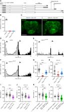
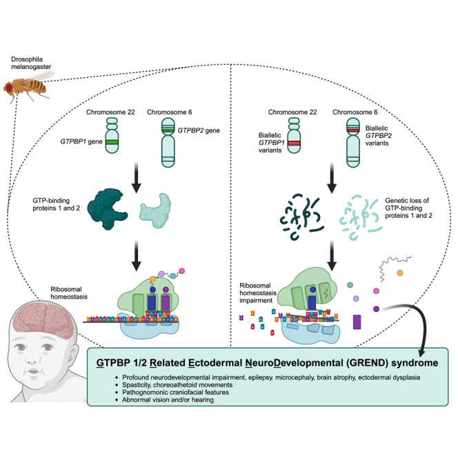
          The homologous genes GTPBP1 and GTPBP2 encode GTP-binding proteins 1 and 2, which are involved in ribosomal homeostasis. Pathogenic variants in GTPBP2 were recently shown to be an ultra-rare cause of neurodegenerative or neurodevelopmental disorders (NDDs). Until now, no human phenotype has been linked to GTPBP1. Here, we describe individuals carrying bi-allelic GTPBP1 variants that display an identical phenotype with GTPBP2 and characterize the overall spectrum of GTP-binding protein (1/2)-related disorders. In this study, 20 individuals from 16 families with distinct NDDs and syndromic facial features were investigated by whole-exome (WES) or whole-genome (WGS) sequencing. To assess the functional impact of the identified genetic variants, semi-quantitative PCR, western blot, and ribosome profiling assays were performed in fibroblasts from affected individuals. We also investigated the effect of reducing expression of CG2017, an ortholog of human GTPBP1/2, in the fruit fly Drosophila melanogaster. Individuals with bi-allelic GTPBP1 or GTPBP2 variants presented with microcephaly, profound neurodevelopmental impairment, pathognomonic craniofacial features, and ectodermal defects. Abnormal vision and/or hearing, progressive spasticity, choreoathetoid movements, refractory epilepsy, and brain atrophy were part of the core phenotype of this syndrome. Cell line studies identified a loss-of-function (LoF) impact of the disease-associated variants but no significant abnormalities on ribosome profiling. Reduced expression of CG2017 isoforms was associated with locomotor impairment in Drosophila. In conclusion, bi-allelic GTPBP1 and GTPBP2 LoF variants cause an identical, distinct neurodevelopmental syndrome. Mutant CG2017 knockout flies display motor impairment, highlighting the conserved role for GTP-binding proteins in CNS development across species.

          Graphical abstract

          Abstract

          Bi-allelic variants in the translational GTPases GTPBP1 and GTPBP2 may affect ribosomal translational control and impair brain development and neurological function. This research identifies 20 individuals with homozygous GTPBP1 and GTPBP2 variants leading to an identical neurodevelopmental syndrome, which we defined as “Gtpbp1/2-related ectodermal neurodevelopmental (GREND) syndrome.”

          Related collections

          Most cited references31

          • Record: found
          • Abstract: found
          • Article: not found

          Standards and Guidelines for the Interpretation of Sequence Variants: A Joint Consensus Recommendation of the American College of Medical Genetics and Genomics and the Association for Molecular Pathology

          The American College of Medical Genetics and Genomics (ACMG) previously developed guidance for the interpretation of sequence variants. 1 In the past decade, sequencing technology has evolved rapidly with the advent of high-throughput next generation sequencing. By adopting and leveraging next generation sequencing, clinical laboratories are now performing an ever increasing catalogue of genetic testing spanning genotyping, single genes, gene panels, exomes, genomes, transcriptomes and epigenetic assays for genetic disorders. By virtue of increased complexity, this paradigm shift in genetic testing has been accompanied by new challenges in sequence interpretation. In this context, the ACMG convened a workgroup in 2013 comprised of representatives from the ACMG, the Association for Molecular Pathology (AMP) and the College of American Pathologists (CAP) to revisit and revise the standards and guidelines for the interpretation of sequence variants. The group consisted of clinical laboratory directors and clinicians. This report represents expert opinion of the workgroup with input from ACMG, AMP and CAP stakeholders. These recommendations primarily apply to the breadth of genetic tests used in clinical laboratories including genotyping, single genes, panels, exomes and genomes. This report recommends the use of specific standard terminology: ‘pathogenic’, ‘likely pathogenic’, ‘uncertain significance’, ‘likely benign’, and ‘benign’ to describe variants identified in Mendelian disorders. Moreover, this recommendation describes a process for classification of variants into these five categories based on criteria using typical types of variant evidence (e.g. population data, computational data, functional data, segregation data, etc.). Because of the increased complexity of analysis and interpretation of clinical genetic testing described in this report, the ACMG strongly recommends that clinical molecular genetic testing should be performed in a CLIA-approved laboratory with results interpreted by a board-certified clinical molecular geneticist or molecular genetic pathologist or equivalent.
            Bookmark
            • Record: found
            • Abstract: found
            • Article: found
            Is Open Access

            GENCODE reference annotation for the human and mouse genomes

            Abstract The accurate identification and description of the genes in the human and mouse genomes is a fundamental requirement for high quality analysis of data informing both genome biology and clinical genomics. Over the last 15 years, the GENCODE consortium has been producing reference quality gene annotations to provide this foundational resource. The GENCODE consortium includes both experimental and computational biology groups who work together to improve and extend the GENCODE gene annotation. Specifically, we generate primary data, create bioinformatics tools and provide analysis to support the work of expert manual gene annotators and automated gene annotation pipelines. In addition, manual and computational annotation workflows use any and all publicly available data and analysis, along with the research literature to identify and characterise gene loci to the highest standard. GENCODE gene annotations are accessible via the Ensembl and UCSC Genome Browsers, the Ensembl FTP site, Ensembl Biomart, Ensembl Perl and REST APIs as well as https://www.gencodegenes.org.
              Bookmark
              • Record: found
              • Abstract: found
              • Article: not found

              Genome-wide analysis in vivo of translation with nucleotide resolution using ribosome profiling.

              Techniques for systematically monitoring protein translation have lagged far behind methods for measuring messenger RNA (mRNA) levels. Here, we present a ribosome-profiling strategy that is based on the deep sequencing of ribosome-protected mRNA fragments and enables genome-wide investigation of translation with subcodon resolution. We used this technique to monitor translation in budding yeast under both rich and starvation conditions. These studies defined the protein sequences being translated and found extensive translational control in both determining absolute protein abundance and responding to environmental stress. We also observed distinct phases during translation that involve a large decrease in ribosome density going from early to late peptide elongation as well as widespread regulated initiation at non-adenine-uracil-guanine (AUG) codons. Ribosome profiling is readily adaptable to other organisms, making high-precision investigation of protein translation experimentally accessible.
                Bookmark

                Author and article information

                Contributors
                Journal
                Am J Hum Genet
                Am J Hum Genet
                American Journal of Human Genetics
                Elsevier
                0002-9297
                1537-6605
                20 December 2023
                04 January 2024
                20 December 2023
                : 111
                : 1
                : 200-210
                Affiliations
                [1 ]Department of Neuromuscular Diseases, UCL Queen Square Institute of Neurology, London, UK
                [2 ]Department of Clinical Genetics, Human Genetics and Genome Research Institute, National Research Centre, Cairo, Egypt
                [3 ]Howard Hughes Medical Institute, Department of Molecular Biology and Genetics, Johns Hopkins University School of Medicine, Baltimore, MD 21205, USA
                [4 ]Molecular Genetics and Functional Genomics, Ospedale Pediatrico Bambino Gesù, IRCCS, 00146 Rome, Italy
                [5 ]Department of Clinical and Experimental Epilepsy, UCL Queen Square Institute of Neurology, London, UK
                [6 ]Institute of Neuro- and Behavioral Biology, Westfälische Wilhelms University, Münster, Germany
                [7 ]Genetic and Precision Medicine Department, King Abdullah Specialized Children Hospital, King Abdulaziz Medical City, Ministry of National Guard Health Affairs (MNGHA), Riyadh, Saudi Arabia
                [8 ]King Abdullah International Medical Research Center (KAIMRC), King Saud bin Abdulaziz University for Health Sciences, Ministry of National Guard Health Affairs (MNGHA), Riyadh, Saudi Arabia
                [9 ]Arcensus GmbH, Rostock, Germany
                [10 ]Division of Medical Genetics, Department of Pediatrics, McGill University, Montreal, Canada
                [11 ]Unit of Medical Genetics, IRCCS Istituto Giannina Gaslini, Genoa, Italy
                [12 ]Department of Neurosciences, Rehabilitation, Ophthalmology, Genetics, Maternal and Child Health, University of Genoa, Genoa, Italy
                [13 ]Unit of Pediatric Neurology, IRCCS Istituto Giannina Gaslini, Genoa, Italy
                [14 ]Department of Medical Genetics, Faculty of Medicine, Mashhad University of Medical Sciences, Mashhad, Iran
                [15 ]Health Biotechnology Division, National Institute for Biotechnology and Genetic Engineering (NIBGE), Faisalabad, Pakistan
                [16 ]National Institute for Health Research Oxford Biomedical Research Centre, Oxford, UK
                [17 ]Genomics Research Centre, Human Technopole, Milan, Italy
                [18 ]Wellcome Centre for Human Genetics, University of Oxford, Oxford OX3 7BN, UK
                [19 ]Town Women and Children Hospital, Peshawar, Pakistan
                [20 ]Neurology Unit, Department of Pediatrics, Faculty of Medicine, Alexandria University, Alexandria, Egypt
                [21 ]Faculty of Medicine, University of Alexandria, Alexandria, Egypt
                [22 ]Department of Family Health, High Institute of Public Health, University of Alexandria, Alexandria, Egypt
                [23 ]Department of Medical Molecular Genetics, Human Genetics and Genome Research Institute, National Research Centre, Cairo, Egypt
                [24 ]3billion, Inc, Seoul, South Korea
                [25 ]Laboratory of Medical Genetics, St. Sophia’s Children’s Hospital, National and Kapodistrian University of Athens, Athens, Greece
                [26 ]Genetics Unit, Department of Obstetrics & Gynaecology, Aristotle University of Thessaloniki, Papageorgiou General Hospital, Thessaloniki, Greece
                [27 ]Paediatric Outpatient Clinic, Xanthi, Greece
                [28 ]Department of Medical Genetics, Faculty of Medicine, Mashhad University of Medical Sciences, Masshad, Iran
                [29 ]Student Research Committee, Faculty of Medicine, Mashhad University of Medical Sciences, Masshad, Iran
                [30 ]Mashhad Genetic Counselling Center, Masshad, Iran
                [31 ]Pediatric Neurology Department, Ghaem Hospital, Mashhad University of Medical Sciences, Mashhad, Iran
                [32 ]Department of Pediatrics, North Khorasan University of Medical Sciences, Bojnurd, Iran
                [33 ]Manchester Centre for Genomic Medicine, St Mary’s Hospital, Manchester University NHS Foundation Trust, Health Innovation Manchester, Manchester M13 9WL, UK
                [34 ]CENTOGENE GmbH, Rostock, Germany
                [35 ]Department of Developmental Neuroscience, IRCCS Stella Maris Foundation, 56128 Pisa, Italy
                [36 ]Department of Clinical and Experimental Medicine, University of Pisa, 56126 Pisa, Italy
                [37 ]Department of Clinical Sciences and Community Health, University of Milan, Milan, Italy
                [38 ]Center for Rare Diseases and Birth Defects, Department of Women and Child Health and Public Health, Fondazione Policlinico Universitario A. Gemelli IRCCS, 00168 Rome, Italy
                [39 ]Department of Pediatrics, University of Chieti, 66100 Chieti, Italy
                [40 ]Unit of Medical Genetics, Children’s Specialist Hospital, King Fahad Medical City, Riyadh, Saudi Arabia
                [41 ]Royal Hospital for Sick Children, Edinburgh, UK
                [42 ]College of Medicine, King Saud Bin Abdulaziz University for Health Sciences, Ministry of National Guard Health Affairs (MNGH), Riyadh, Saudi Arabia
                [43 ]Department of Genetics, King Faisal Specialist Hospital and Research Center, Riyadh, Saudi Arabia
                [44 ]Department of Neuroradiology, Great Ormond Street Hospital, London, UK
                [45 ]Genetics Research Centre, Molecular and Clinical Sciences Institute, University of London, St George’s, Cranmer Terrace, London SW17 0RE, UK
                [46 ]Department of Pediatrics, Boston Children’s Hospital Harvard Medical School, Boston, MA 02115, USA
                Author notes
                []Corresponding author h.houlden@ 123456ucl.ac.uk
                [47]

                These authors contributed equally

                Article
                S0002-9297(23)00430-5
                10.1016/j.ajhg.2023.11.012
                10806450
                bd21855f-b786-444b-bbdb-e5909ecb8f0d
                © 2023 The Authors

                This is an open access article under the CC BY license (http://creativecommons.org/licenses/by/4.0/).

                History
                : 15 May 2023
                : 29 November 2023
                Categories
                Report

                Genetics
                neurodevelopmental disorders,neurodegeneration,nbia,ribosomopathies,ribosome stalling,gtpbp1,gtpbp2,animal models,ectodermal disorders,grend syndrome

                Comments

                Comment on this article