18
views
0
recommends
+1 Recommend
0 collections
    0
    shares
      • Record: found
      • Abstract: found
      • Article: found
      Is Open Access
      research-article

      Read this article at

      Bookmark
          There is no author summary for this article yet. Authors can add summaries to their articles on ScienceOpen to make them more accessible to a non-specialist audience.

          Summary

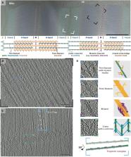
          Sarcomeres are force-generating and load-bearing devices of muscles. A precise molecular picture of how sarcomeres are built underpins understanding their role in health and disease. Here, we determine the molecular architecture of native vertebrate skeletal sarcomeres by electron cryo-tomography. Our reconstruction reveals molecular details of the three-dimensional organization and interaction of actin and myosin in the A-band, I-band, and Z-disc and demonstrates that α-actinin cross-links antiparallel actin filaments by forming doublets with 6-nm spacing. Structures of myosin, tropomyosin, and actin at ~10 Å further reveal two conformations of the “double-head” myosin, where the flexible orientation of the lever arm and light chains enable myosin not only to interact with the same actin filament, but also to split between two actin filaments. Our results provide unexpected insights into the fundamental organization of vertebrate skeletal muscle and serve as a strong foundation for future investigations of muscle diseases.

          Graphical abstract

          Highlights

          • Three-dimensional sarcomere organization and plasticity at the molecular level

          • Myosin double heads can adopt two different interactions with actin filaments

          • Transition between tropomyosin states happens within one tropomyosin unit

          • An irregular mesh of α-actinin doublets cross-links antiparallel actin filaments

          Abstract

          Visualizing the mouse sarcomere in the rigor state using electron cryo-tomography reveals architectural details of the different zones and provides insight into how key factors are arranged within them to support function during muscle contraction.

          Related collections

          Most cited references131

          • Record: found
          • Abstract: found
          • Article: not found

          Fiji: an open-source platform for biological-image analysis.

          Fiji is a distribution of the popular open-source software ImageJ focused on biological-image analysis. Fiji uses modern software engineering practices to combine powerful software libraries with a broad range of scripting languages to enable rapid prototyping of image-processing algorithms. Fiji facilitates the transformation of new algorithms into ImageJ plugins that can be shared with end users through an integrated update system. We propose Fiji as a platform for productive collaboration between computer science and biology research communities.
            Bookmark
            • Record: found
            • Abstract: found
            • Article: not found

            NIH Image to ImageJ: 25 years of image analysis

            For the past twenty five years the NIH family of imaging software, NIH Image and ImageJ have been pioneers as open tools for scientific image analysis. We discuss the origins, challenges and solutions of these two programs, and how their history can serve to advise and inform other software projects.
              Bookmark
              • Record: found
              • Abstract: found
              • Article: not found

              UCSF Chimera--a visualization system for exploratory research and analysis.

              The design, implementation, and capabilities of an extensible visualization system, UCSF Chimera, are discussed. Chimera is segmented into a core that provides basic services and visualization, and extensions that provide most higher level functionality. This architecture ensures that the extension mechanism satisfies the demands of outside developers who wish to incorporate new features. Two unusual extensions are presented: Multiscale, which adds the ability to visualize large-scale molecular assemblies such as viral coats, and Collaboratory, which allows researchers to share a Chimera session interactively despite being at separate locales. Other extensions include Multalign Viewer, for showing multiple sequence alignments and associated structures; ViewDock, for screening docked ligand orientations; Movie, for replaying molecular dynamics trajectories; and Volume Viewer, for display and analysis of volumetric data. A discussion of the usage of Chimera in real-world situations is given, along with anticipated future directions. Chimera includes full user documentation, is free to academic and nonprofit users, and is available for Microsoft Windows, Linux, Apple Mac OS X, SGI IRIX, and HP Tru64 Unix from http://www.cgl.ucsf.edu/chimera/. Copyright 2004 Wiley Periodicals, Inc.
                Bookmark

                Author and article information

                Contributors
                Journal
                Cell
                Cell
                Cell
                Cell Press
                0092-8674
                1097-4172
                15 April 2021
                15 April 2021
                : 184
                : 8
                : 2135-2150.e13
                Affiliations
                [1 ]Department of Structural Biochemistry, Max Planck Institute of Molecular Physiology, Otto-Hahn-Strasse 11, 44227 Dortmund, Germany
                [2 ]The Randall Centre for Cell and Molecular Biophysics, School of Basic and Medical Biosciences, Kings College London BHF Excellence Centre, New Hunt’s House, Guy’s Campus, London SE1 1UL, UK
                Author notes
                []Corresponding author stefan.raunser@ 123456mpi-dortmund.mpg.de
                [3]

                These authors contributed equally

                [4]

                Lead contact

                Article
                S0092-8674(21)00236-1
                10.1016/j.cell.2021.02.047
                8054911
                33765442
                2bbd79c4-c51d-405c-8ba8-d15fcefba0b9
                © 2021 The Author(s)

                This is an open access article under the CC BY license (http://creativecommons.org/licenses/by/4.0/).

                History
                : 12 October 2020
                : 27 January 2021
                : 22 February 2021
                Categories
                Article

                Cell biology
                muscle,sarcomere,actin,myosin,z-disc,tropomyosin,electron tomography,structure
                Cell biology
                muscle, sarcomere, actin, myosin, z-disc, tropomyosin, electron tomography, structure

                Comments

                Comment on this article